Certified under IVDR
Standardizing hot spot analysis
Cancer can rapidly mutate and adapt, potentially producing heterogeneous tumors making it important to evaluate a specimen in a hot spot, where the tumor cells have increased activity.
Hot spot analysis for Ki-67 scoring has often been used for proliferation index assessment in breast cancer [1]. However, manual hot spot scoring is subjective and prone to both intra- and inter-observer variability [2]. If not accounted for, non-invasive cancer structures can also affect both hot spot location and proliferation index.
The Hot Spot APP provides an automatic and objective hot spot scoring that has been shown to outperform manual biomarker assessment [3]. The APP presents pathologists with a heatmap for easy visualization of tumor heterogeneity.
Configurable for your analysis
The Hot Spot APP is configurable for both shape and size to facilitate analysis of different types of tissue and stain. To the right, four hot spot examples are shown with corresponding proliferation indices. The Hot Spot APP facilitates both a predefined size and shape or an adaptable hot spot with a predefined number of nuclei.
Complete Ki-67 analysis workflow
The Hot Spot APP works in a fully automatic workflow
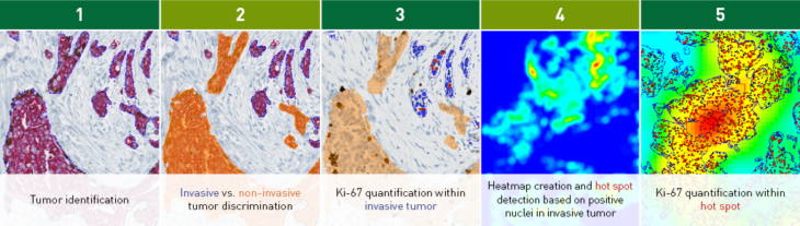
The workflow example above considers a Ki-67 IHC analysis. Tumor is automatically identified and separated into invasive and non-invasive regions based on a physical double stain of p63+CK7/19 (Step 1+2). Ki-67 is quantified within the invasive tumor components (Step 3).
The Hot Spot APP creates a heatmap based on the density of the positive nuclei (Step 4), identifies a hot spot, and outputs the Ki-67 proliferation index (%) for the hot spot (Step 5).
In EU/UK: CE IVD – for use in diagnostic procedures