Developed for tumor detection in H&E stained prostate tissue
Prostate cancer is the second most common cancer in men, with an estimated 1.1 million diagnoses worldwide in 2012, accounting for 15% of all cancers diagnosed [1]. Research in prostate cancer is important to help improve diagnosis and choosing the best treatments for individuals at all stages of the disease. Automated tumor detection can help identify regions-of-interest and provide numerical data in a scalable fashion.
This APP utilizes AI/deep learning and has been trained to detect tumors in images of prostate tissue stained with H&E. The deep learning architecture enables the APP to recognize complex structures and interpret the tissue context when analyzing an image, making it an efficient tool for detecting even small tumors that are not easily noticed. The APP does not grade the tumors.
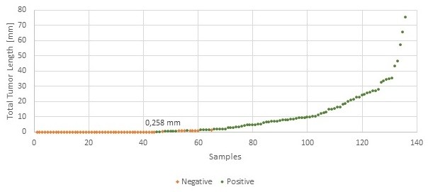
136 slides were manually assessed and classified as positive or negative, resulting in 53 negative and 83 positive slides. The same slides were analyzed with the APP where the Total Tumor Length with a cut-off of 0 mm was used to classify slides as positive and negative. The agreement between manual and APP slide classification is shown below.
| Sensitivity | 97.9 % |
|---|---|
| Specificity | 83.0 % |