Author: Bettina Winkler

  • Minerva Imaging and Visiopharm announce collaboration on AI-based image analysis

    Minerva Imaging and Visiopharm announce collaboration on AI-based image analysis

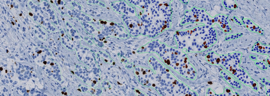
    Visiopharm and Minerva Imaging have announced a collaboration on development of sophisticated AI-based image analysis applications in histology. Visiopharm’s AI-driven Oncotopix® platform has the potential to play a crucial role in developing powerful new predictive assays. The main focus within Minerva Imaging will be on targeted radionuclide therapy and immuno-oncology, enabling to gauge the effectiveness of interventions and making data-driven decisions.

    Targeted radionuclide therapy

    Targeted radionuclide therapy is a form of precision medicine where a radioactive substance is attached to a targeting moiety. It is administered through the body to specifically target cancer cells and irradiate them. The efficacy of these treatments can be determined by the level of radio-induced DNA damage that it causes. In one of the first cooperative projects Minerva and Visiopharm will develop quantitative AI-based image analysis to assess this level of DNA damage.

    Carsten H. Nielsen, CEO and co-founder of Minerva Imaging, said:

    “Targeted radionuclide therapy has gained a lot of momentum in recent years in the oncology area. We are convinced that the development of powerful and highly accurate AI-based image analysis for determining the level of DNA damage and other aspects of therapeutic efficacy will be very valuable. We are committed to bringing this and a number of other AI-based applications to the market impacting the advancement of new, improved, and lifesaving drugs for seriously ill cancer patients.”

    Michael Grunkin, co-founder and CEO of Visiopharm, said:

    “We are excited to establish this strategic collaboration with Minerva to accelerate the development of new AI-based histology analysis in oncology. This collaboration will allow us to support an increasingly important precision medicine agenda.”

    AI-driven image analysis

    AI-driven precision pathology has the potential to play a crucial role in developing powerful new predictive assays. It provides a powerful tool for discovery of new biomarkers through the analysis of multiplexed image data, developing investigational devices for clinical trials, and developing scoring principles for novel tissue biomarkers. Moreover, it can aid in the development of image analysis for standardized and automated reading of increasingly complex biomarkers.

    The Minerva-Visiopharm partnership will speed up the development of a toolbox for AI-driven precision pathology, enabling Pharma to accelerate clinical development by improving quantitative and qualitative assessments in hard-to-treat cancers.

    About Minerva Imaging

    Minerva Imaging is a scientifically driven CRO founded in 2011, and currently counting over 130 employees. The company focuses on the use of advanced models and molecular imaging for translational research and drug development. Minerva Imaging engages with its clients to understand their scientific questions and discuss how its methods and capabilities can provide answers. Minerva worked with over 100 global pharma and biotech companies.

    Follow us on LinkedIn for the latest news updates or visit www.minervaimaging.com for more information.

    About Visiopharm

    Visiopharm® is a leading provider of AI-driven precision pathology software for research and diagnostics. In research, it is a technology leader providing tools that help scientists, pathologists, and image analysis experts produce accurate data for all types of tissue-based research. In diagnostics, it is a leader within clinical applications, with no less than eight diagnostic algorithms cleared under IVDR for EU and UK customers. These applications provide diagnostic decision support and can be easily activated and integrated into existing lab workflows.

    Founded in 2002, Visiopharm is privately owned and operates internationally with over 750 customer accounts in more than 40 countries. The company’s headquarters are located in Denmark’s Medicon Valley, with offices in Sweden, the UK, Germany, the Netherlands, and the United States, and local representation in France and China.

    Media enquiries

    Eske Rygaard-Hjalsted
    Senior Business Developer
    erh@minervaimaging.com
    +45 20 30 22 36

    Johanne Louise Brændgaard
    Chief Marketing Officer
    jlb@visiopharm.com

  • Utilization of Deep Learning is Growing in Digital Pathology

    ​Digital Pathology is undergoing a revolutionary change with the introduction of state-of-the-art pattern recognition via/with deep learning. The transition from feature engineering and classical machine learning to end-to-end deep learning has happened at a rapid pace. Some of this fast adoption rate can be attributed to the improved results that deep learning can provide to pathologists while its use as a decision support tool in case management organization is also a driver on the horizon.

    In addition, as Graphical Processing Units (GPUs) gain more computational power, the parallelization required for execution and training of deep learning algorithms continues to improve. This increased performance and decrease in cost of these once expensive “gaming” GPUs have opened a large market for deep learning in digital pathology.

    ​​Deep learning is more than a classifier


    Based on the adoption of deep learning one might be inclined to think that deep learning is just another specific type of classifier such as a Random Forest or K-Nearest Neighbor which have specific rulesets for functionality. However, deep learning is a whole scientific field and there are many different implementations of deep learning frameworks and architectures which can yield a spectrum of results from poor to excellent. When choosing a provider of deep learning, you should be assured that the neural network architecture they are using is state-of-the-art as the field has moved extremely fast in the last several years. 

    Visiopharm has taken the approach of including multiple networks for deep learning in our AI Deep Learning package. The reason for this inclusion is that some deep learning networks are good for one bucket of tasks while other networks are good for a second bucket of tasks and so on. Limiting the user’s choice to a single network is essentially limiting their work to a single bucket and thus reducing the places deep learning can be effective for their science. 

    ​Flexibility in deep learning is required for optimal performance


    Deep Convolutional (or Neural) Networks themselves also have architectures that can be modified so that deep learning is optimized for a given image analysis task, but this requires intimate knowledge of deep learning and its implementations in Python or another programming language. Visiopharm always prides itself on providing its customers with a highly configurable toolbox and deep learning architectures have been added to this list of customizations with the release of AI Architect. 

    AI Architect is an advanced module that lets the user optimize deep learning networks for their needs. This toolbox allows users to enhance the performance of deep learning, speed up deep learning and provides solutions for training when data sources are limited. The best part about the AI Architect: no programming knowledge necessary! The user gets an intuitive GUI from within our AI Deep Learning Author that provides the user with an easy to use suite of settings to adjust AI network architecture to suit their scientific needs. 

    So, whether you have previous practical experience with deep learning, just knowing the basics, or just starting to consider using deep learning in your research, you can use our state-of-the-art deep learning-based platform in your research! 

    Contact Visiopharm today for a demo!

  • OracleBio adds Visiopharm Oncotopix software for Image Analysis

    OracleBio, a global leader in high precision, quantitative digital image analysis services, is pleased to announce an expansion of its software capabilities to include Visiopharm Oncotopix®. Produced by Denmark-based Visiopharm, Oncotopix® software is an advanced platform for computer assisted quantification of immunohistochemistry (IHC) and In-Situ Hybridization (ISH) staining. Oncotopix offers a portfolio of software modules designed for application within cancer diagnostics and for scientists within cancer research and drug development.

    Lorcan Sherry, Chief Scientific Officer of OracleBio commented:

    ‘It is important that OracleBio continues to develop and deliver highly innovative histology image analysis services to support our Client’s pre-clinical and clinical R&D. Visiopharm Oncotopix is highly interactive and infinitely programmable, allowing OracleBio to custom configure image analysis requirements and define new algorithms to meet Clients’ specific requirements.’

    Whilst Visiopharm offers a range of different solutions for digital pathology, the Oncotopix platform in particular will strengthen OracleBio’s Immuno-Oncology service offering. The software provides key features that support oncology research, such as virtual multiplexing, and is a powerful tool for advanced co-localisation studies and tumor detection.

    Lars Pedersen, Director of Sales at Visiopharm, commented:

    “Visiopharm is proud to include OracleBio in our growing base of pre-clinical and clinical CRO clients. The Oncotopix® solution that OracleBio has chosen is designed to match the increasing demand for high quality tissue-based biomarker analysis, shorter turn-around times and flexibility, and we are proud to fuel OracleBio in their ambitions to deliver innovative expert histopathology results with our proprietary solution for in vitro diagnostic applications.”

    Along with OracleBio’s current analysis platform capabilities the addition of Visiopharm Oncotopix will further enhance the company’s service capabilities and puts OracleBio in the unique position of being a multi-image analysis platform service provider.