In a groundbreaking collaboration, Visiopharm and the UK National External Quality Assessment Scheme for Immunocytochemistry and In-Situ Hybridisation (UK NEQAS ICC & ISH) are setting new standards in the quality control of IHC testing, harnessing cutting-edge AI technology alongside rigorous external quality assessment.
The need for this initiative is clearly demonstrated in the highly concerning data that reveal 25-30% of laboratories repeatedly fail to meet minimum proficiency standards when testing for critical predictive biomarkers, most often due to false negative results. Given that these tests significantly influence patient treatment decisions, the implications of such inaccuracies are profound.
The alliance introduces a comprehensive toolkit for pathology laboratories, designed to maintain staining quality and align with the latest clinical and regulatory requirements. Visiopharm’s innovative Qualitopix solution enables daily and continuous measuring, monitoring, and documentation of staining consistency/quality, promptly identifying and addressing any deviations. When coupled with UK NEQAS ICC & ISH’s rigorous four-monthly assessments, the laboratory is assured of end-to-end quality assurance monitoring.
Mr. Andy Dodson, Director of UK NEQAS ICC & ISH, highlighted the gravity of the current situation: “The present figures concerning IHC testing accuracy are worrying. The direct link between test precision and patient outcomes cannot be overstated. This joint initiative, combining Visiopharm’s AI-driven internal quality control with our external quality assessment, aims to significantly enhance testing quality, a critical factor in delivering optimal patient care.”
This initiative is poised to redefine industry benchmarks, facilitating laboratories’ adherence to the stringent requirements of the recently revised ISO15189:2022 standard and other regulatory mandates, thereby ensuring superior patient care.
Dr. Michael Grunkin, CEO of Visiopharm, stressed the initiative’s ultimate goal: “At the heart of our mission is patient care. Through this collaboration, we are committed to providing laboratories with the advanced tools necessary for enhancing diagnostic precision. Our joint efforts are expected to play a pivotal role in ensuring patients receive the most effective treatments available.“
About Visiopharm
Visiopharm is a leading provider of AI-driven precision pathology software for research and diagnostics. In research, it is a technology leader providing tools that help scientists, pathologists, and image analysis experts produce accurate data for all types of tissue-based research. In diagnostics, it is a leader within clinical applications, with no less than nine diagnostic algorithms cleared under IVDR for EU and UK customers. These applications provide diagnostic decision support and can be easily activated and integrated into existing lab workflows. Founded in 2002, Visiopharm is privately owned and operates internationally with over 750 customer accounts in more than 40 countries. The company’s headquarters are located in Denmark’s Medicon Valley, with legal entities in Sweden, the UK, Germany, the Netherlands, and the United States, and local representation in France and China.
UK NEQAS ICC & ISH is at the forefront of external quality assessment for advanced diagnostic testing in cellular pathology, committed to elevating the standards of IHC and ISH biomarker testing and ensuring adherence to the highest quality and regulatory standards. Founded in 1986, it was one of the first to introduce external quality assessment into clinical cellular pathology. The Scheme is fully accredited to ISO/IEC standards both in the UK and internationally. It operates worldwide, providing a fully comprehensive EQA service to clinical laboratories in more than 50 countries.
CND’s Biomarker Testing Services Utilizing Groundbreaking NerValence™ System Unveiled at the AD/PD™ 2024 Parkinson’s and Alzheimer’s Disease Conference
Scottsdale, Ariz – CND Life Sciences (CND), a medical technology company pioneering the development of cutaneous neurodiagnostic tests and associated biomarker services, today will unveil its NerValence pathology detection system at the AD/PD 2024 conference in Lisbon, Portugal. NerValence, developed through a strategic collaboration with precision pathology company Visiopharm, significantly enhances CND’s biomarker testing services for clinical trials targeting the alpha-synuclein protein in patients with suspected Parkinson’s disease, dementia with Lewy bodies, and related disorders.
“As an expansion of CND’s cutaneous testing services, NerValence is poised to transform the way we identify, quantify, and monitor the misfolded, pathological form of the alpha-synuclein protein in patients,” said Todd Levine, MD, CND’s Chief Medical Officer. “Our work with Visiopharm has demonstrated how innovative digital technology can enhance the detection and measurement of this key biomarker to optimize drug development for Parkinson’s disease and other synuclein-based disorders.”
NerValence features machine learning algorithms that recognize and differentiate microscopic structures, nerves, and important disease markers that can be found in cutaneous tissue. The system was designed and developed by CND in partnership with Visiopharm as a pathology assistance tool for CND’s Syn-One Test® and is used exclusively by CND pathologists and researchers at the company’s CLIA-certified laboratories in Scottsdale. Syn-One relies on the analysis of skin biopsy samples collected from patients who have signs and symptoms of Parkinson’s disease and related disorders known as synucleinopathies and has demonstrated greater than 95% sensitivity and specificity in detecting phosphorylated alpha-synuclein. Syn-One, as supported by NerValence, is a laboratory developed test (LDT) ordered by neurologists and other clinicians across the US to aid the diagnosis of thousands of patients. NerValence’s unique ability to enhance the identification, tracking, and measuring the volume of alpha-synuclein in cutaneous nerves offers significant benefit in targeting clinical trial patients for the development of disease-modifying therapies.
“Utilizing our novel deep learning technology to power this crucial advancement in neurodegenerative diseases represents a significant leap forward in analyzing peripheral nerves,” said Michael Grunkin, Visiopharm Chief Executive Officer. “Collaborating with CND Life Sciences on its deployment of NerValence will continue to be a major priority as we advance AI-driven precision pathology to enhance biopharmaceutical drug development.”
Stop by our Exhibit Booth #66 at the AD/PD 2024 conference to get a first-hand look at the NerValence system. Also be sure to check out our posters titled “The Synuclein-One Study: Cutaneous Alpha-Synuclein Deposition in Parkinson’s Disease,” Poster Board #0544 and “The Synuclein-One Study: Cutaneous Alpha-Synuclein Deposition in Dementia with Lewy Bodies and Mild Cognitive Impairment,” Poster Board #0561. Both posters will be available for viewing from March 6-7 during designated poster hours.
About CND Life Sciences
CND Life Sciences is dedicated to supporting the care of patients facing the potential diagnosis of a neurodegenerative disease and other neurological conditions. Operating a CLIA-certified and CAP-accredited laboratory in Scottsdale, Arizona, CND launched the Syn-One Test® in 2019 as the world’s first commercially available test to detect, visualize, and quantify phosphorylated alpha-synuclein located in cutaneous nerves. This laboratory developed test analyzes three small skin biopsies collected from the patient in a physician’s office through a 15-minute minimally invasive procedure and includes an assessment of intraepidermal nerve fiber density and other important pathologic evaluations in the skin. The Syn-One Test is supported by technology licensed from Beth Israel Deaconess Medical Center in Boston and leverages a decade of published science from leading academic institutions in multiple countries. The Company has research collaborations with multiple biopharmaceutical companies and has been awarded three prestigious NIH SBIR grants to advance the validation and clinical utility of its Syn-One Test. For more information visit cndlifesciences.com or connect with us on LinkedIn.
Disclosure: Research reported in this publication was supported by the National Institute of Neurological Disorders and Stroke and the National Institute on Aging (NIA) of the National Institutes of Health under Award Numbers R44NS117214, R44AG076072, and R44NS127696. The content is solely the responsibility of the authors and does not necessarily represent the official views of the National Institutes of Health.
About Visiopharm
Visiopharm is a leading provider of AI-driven precision pathology software for research and diagnostics. In research, it is a technology leader providing tools that help scientists, pathologists, and image analysis experts produce accurate data for all types of tissue-based research. In diagnostics, it is a leader within clinical applications, with no less than nine diagnostic algorithms cleared under IVDR for EU and UK customers. These applications provide diagnostic decision support and can be easily activated and integrated into existing lab workflows. Founded in 2002, Visiopharm is privately owned and operates internationally with over 750 customer accounts in more than 40 countries. The Company’s headquarters are located in Denmark’s Medicon Valley, with legal entities in Sweden, the UK, Germany, the Netherlands, and the United States, and local representation in France and China. For more information visit visiopharm.com.
Exciting news for the world of science and digital pathology!
Colorado State University (CSU), globally recognized research and veterinary institution, in collaboration with Visiopharm, a leader in image analysis software, is thrilled to announce its partnership in a graduate course in digital pathology. This innovative program will teach 42 graduate students modern digital methods in pathology practice, emphasizing the research and clinical applications of digital pathology (DP) tools.
This course is a unique collaboration featuring lectures and platform training by Visiopharm, complemented by the instruction of Dr. Mac Harris and Dr. Brendan Podell, assistant professors in the CSU Department of Microbiology, Immunology and Pathology, and seasoned Visiopharm users.
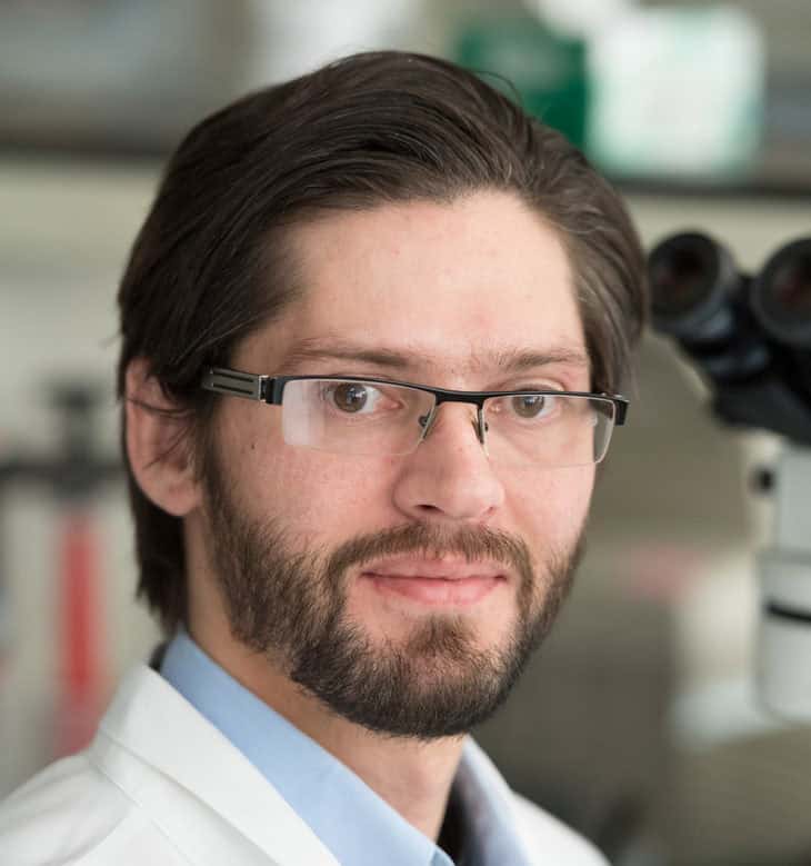
Dr. Mac Harris, DVM, PhD, DACVP Assistant Professor, Microbiology, Immunology, and Pathology
Dr. Brendan Podell, DVM, PhD, DACVP Assistant Professor, Mycobacteria Research Laboratories
A key component of the course involves hands-on experience, where students will embark on an independent project using Visiopharm. These projects, crucial for practical understanding, will be presented between weeks 7 to 9 of the spring semester.
The primary audience for this course comprises graduate students, predominantly from Veterinary Science and Animal Science backgrounds. These bright minds are the future of independent research labs and Veterinary Pathology research, likely to make significant contributions in the next 2-3 years.
Stay tuned for more updates, interviews, and insights as we embark on this exciting journey of innovation and learning.
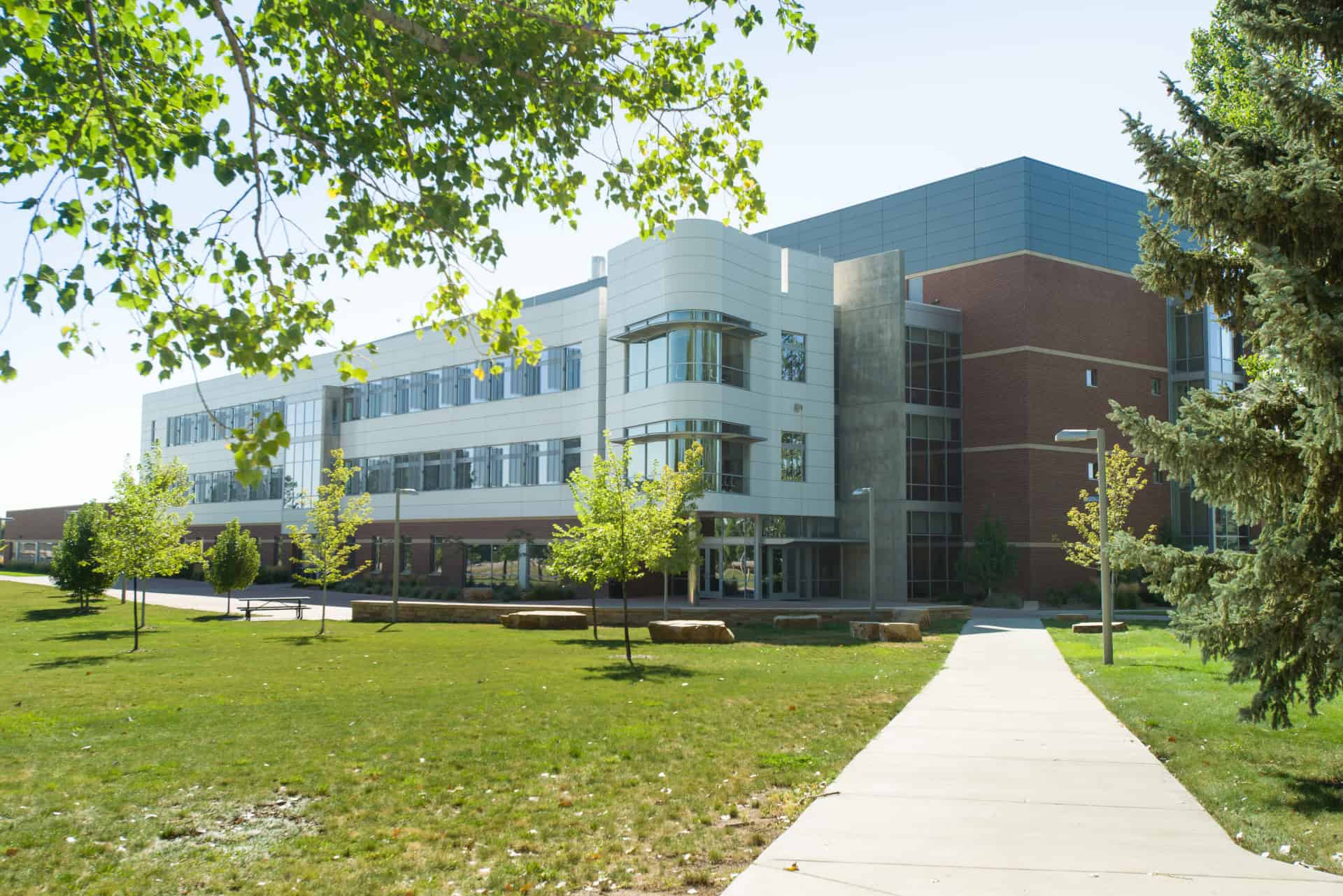
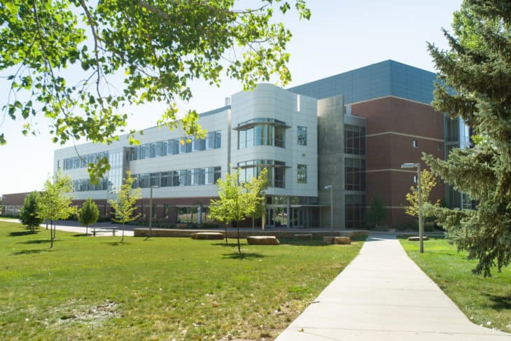
College of Veterinary Medicine and Biomedical Sciences. Colorado State University.
In today’s blog, dive into the engaging Q&A session with OracleBio, a pioneering CRO in quantitative digital pathology, as they discuss their journey with Visiopharm’s software.
The interview with Gabriel Reines March (R&D Project Manager at OracleBio) and Karen McClymont (Image Analysis Project Manager at OracleBio) offers an insight into how OracleBio leverages cutting-edge image analysis and AI technologies to enhance research and development in pharma and biotech globally. From innovative algorithm development to their transition to a cloud-based infrastructure, discover the pivotal role Visiopharm plays in OracleBio’s work, especially in the fields of oncology and immuno-oncology.
Gabriel Reines March R&D Project Manager
With a PhD in Biomedical Image Processing and a background in Electrical Engineering, Gabriel is the OracleBio R&D team lead. He oversees and manages the group’s project pipeline and ensures that the company stays at the bleeding edge of the industry.
Gabriel also manages OracleBio’s involvement in the INCISE project – a collaborative effort between industry, academia and the UK’s National Health Service.
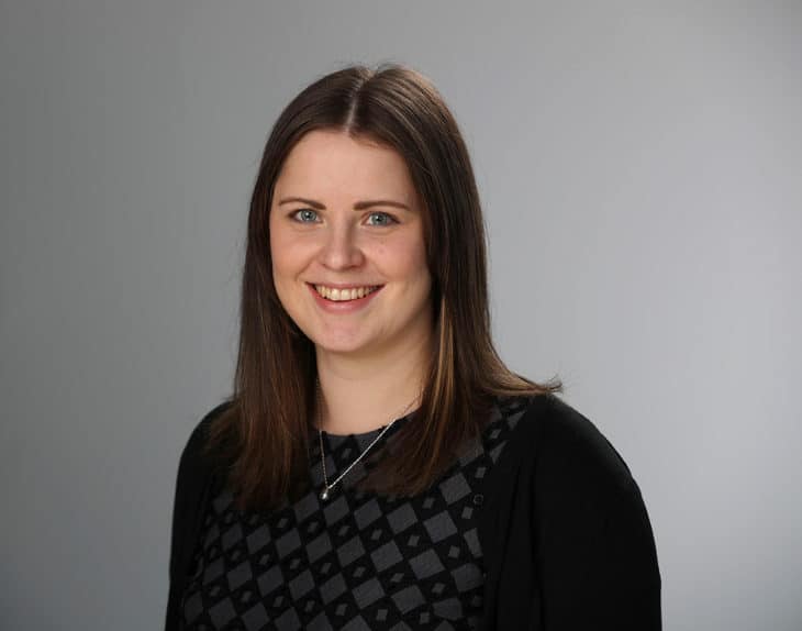
Karen McClymont Image Analysis Project Manager
With a PhD in Biochemistry and her active involvement and support in the analysis of some of OracleBio’s most complex studies, Karen plays a key role leading OracleBio’s image analysis projects and overseeing the team’s continued development.
Visiopharm: Can you tell me a bit about OracleBio and which services you are offering to your customers?
OracleBio: OracleBio is a leading Contract Research Organization (CRO) that provides quantitative digital pathology services to Pharma and Biotech globally. We help our customers by generating accurate and robust data from their histology images, enabling confident interpretation and efficient decision making for their R&D projects.
As well as experienced image analysts, our team is also made up of clinical pathologists, software engineers, and Pharma scientists. This broad expertise base allows us to advise our clients on the best histology staining and analysis approach to ensure relevant image analysis data can be generated in the most optimal manner.
We utilise commercially available image analysis software like Visiopharm and support all stages of the R&D pipeline, from target discovery to clinical trials, with a distinct edge in oncology, immuno-oncology, and fibrosis. Our expertise extends to a diverse range of histological techniques, such as H&E, Immunohistochemistry, Multiplex Immunofluorescence, in situ Hybridization, and Imaging Mass Cytometry. The power and versatility of the software, with the addition of deep learning capabilities, is well-suited to the innovative way OracleBio approaches algorithm development for the multitude of staining profiles that we encounter.
Important to the efficient delivery of data is our image analysis workflow, hosted on our purpose-built, state-of-the-art Amazon Web Services (AWS) cloud that is designed to enable quantitative digital pathology workflows at speed and scale. What’s more, in order to facilitate client engagement and collaboration throughout the study’s lifecycle, we have created our own Visiopharm-powered remote image viewing portal, OBserver, where clients can log on to generate annotations and review analysis overlays.
The power and versatility of the software, with the addition of deep learning capabilities, is well-suited to the innovative way OracleBio approaches algorithm development for the multitude of staining profiles that we encounter.
Visiopharm: Since when have you been using Visiopharm and what led you to Visiopharm originally?
OracleBio: As a Digital Pathology CRO, our primary focus is to generate robust data from our clients’ images. Having a powerful and flexible image analysis software such as Visiopharm provides us with options for innovative solutions to the more challenging image analysis problems. We integrated Visiopharm into our roster of image analysis tools back in 2016. Some of the software features that attracted us were:
the inclusion of different Deep Learning neural networks to train and deploy tissue classifier and cell segmentation apps;
the availability of a comprehensive collection of image post-processing steps, useful for customizing apps to suit specific study requirements;
the flexibility around app development for building bespoke image analysis workflows.
For these reasons, Visiopharm quickly became an integral part of our image analysis workflow.
Visiopharm: Can you describe the process of implementing Visiopharm and how your experience has been working with our team?
OracleBio: When we first introduced Visiopharm into OracleBio’s image analysis workflow, the software was run locally on our on-premise server, which meant we only had access to two user licenses and finite hardware resources. Although this model suited our needs at the time, the limitations of such a rigid IT infrastructure soon became apparent.
As the company expanded, so did the number of concurrent Visiopharm users. In parallel, developments in Artificial Intelligence (AI) powered image analysis technologies translated into an increased demand for GPU-driven Deep Learning algorithms. In response to this, we migrated our IT infrastructure to a purpose-built AWS cloud environment, with flexibility, scalability and speed at the heart of it. Our proprietary ‘QDPConnect’ cloud management system, built with our sister company Sciento, enables our Image Analysis scientists to launch virtual machines containing Visiopharm, with the desired hardware specifications such as GPU support, processing power and memory, to suit specific study requirements.
It’s always a great experience to interact with the Visiopharm team! Their technical experts are always on hand to answer any questions, from supporting installation of the latest software update, to offering advice on specific software issues. A number of our team have also benefited from attending Visiopharm Academy courses to further develop their knowledge of the software. More recently, we joined Visiopharm’s Beta-Tester partner programme, where we get access to the latest software releases and provide expert feedback on the new implemented features and tools, such as the Phenoplex module and the Guided QC workflow.
It’s always a great experience to interact with the Visiopharm team! Their technical experts are always on hand to answer any questions, from supporting installation of the latest software update, to offering advice on specific software issues.
Visiopharm: What kind of problems or questions of your customers can you address with Visiopharm?
OracleBio: We use Visiopharm on a daily basis to provide answers to the research questions posed by our customers. This can range from a simple tumour-stroma classifier on a single-plex chromogenic IHC study, to more complex cell detection and phenotyping on high-plex immunofluorescence assays.
The Tissuealign™ module within Visiopharm has been very valuable when clients have requested segmentation of the tumour microenvironment of IHC and IF images in the absence of a specific tumour marker or any distinctive tumour morphology. Co-registration of the stained tissue images with their corresponding H&Es has enabled us to establish more accurate segmentation which has subsequently led to the generation of more robust data.
Our clients are often interested in the downstream application of bioinformatics. The flexibility in custom-built apps allows us to generate a large amount of useful information defined by the output variables; from a simple cell count (including positive and negative cell populations) to specific x-y coordinates for each cell and mean intensity data for all marker layers present. The exported data can be compiled into formats compatible with a client’s bioinformatics platform for subsequent data analysis.
Visiopharm: What specific features of our product do you find most helpful?
OracleBio: The flexibility in app development is one of the most useful features of Visiopharm and allows us to undertake a variety of projects ranging from single-plex IHC to challenging multiplex IF analysis. Being able to custom-build algorithms to address image analysis challenges is a major advantage of the software. There are a number of routes to take during algorithm development and depending on the tissue type, marker staining and required outputs, algorithms can be easily adapted to optimise the results for each study.
The flexibility in app development is one of the most useful features of Visiopharm and allows us to undertake a variety of projects ranging from single-plex IHC to challenging multiplex IF analysis.
More specifically, the addition of AI to the software has greatly improved our experience, especially with regards to nuclei segmentation. Variable stain intensity is a common factor seen within and between batches of samples, predominantly in clinical studies. The implementation of AI tools has allowed us to develop more robust algorithms that can be applied across a greater range of staining and tissue types. Additionally, the continual software updates, which have recently included improvement to marker visualization and the usability of the colour adjustment tools, provide an enhanced overall user experience.
Visiopharm: Can you share any recent results/outcomes that you have achieved using the platform?
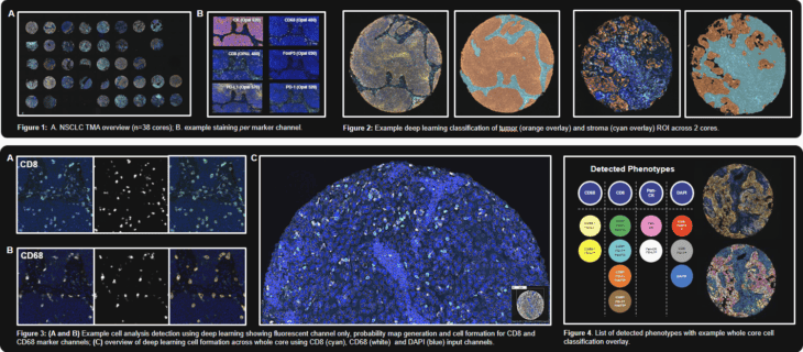
OracleBio: Last year, our collaborative poster with Akoya Biosciences was presented at AACR 2023. OracleBio performed image analysis on samples stained with one of Akoya’s signature mIF panels. We used Visiopharm to profile the immune contexture and tumour spatial interactions in a set of tissue cores, covering a range of NSCLC subtypes and tumour staging. Our approach started with the generation of CD8, CD68 and DAPI cell objects using a Deep Learning algorithm trained on a range of TMA cores. A hierarchical post processing approach was then applied to create CD8, CD68, PanCK and remaining DAPI cell objects for downstream phenotyping. We generated cell object data per core using Visiopharm and carried out spatial analysis using an OracleBio proprietary program (PhenoXplore) to perform neighbourhood analysis for selected phenotypes.
For more details on how we’ve used Visiopharm to analyse samples by incorporating a hierarchal approach to cell detection and phenotyping, check out this case study where we analysed 8-plex multiplex IF staining in CRC & NSCLC tissue. Tissue segmentation using AI deep learning is also demonstrated within Pathology-defined tumour microenvironment regions.
In this this case study, the OracleBio team used Phenoplex to generate phenotyping data of an 8-plex multiplex IF staining by Ultivue in CRC and NSCLC tissues.
Another poster, presented at the Digital Pathology & AI Congress US 2022, demonstrates how we utilised Visiopharm to develop image analysis techniques to quantify HBV on liver samples. The study samples were stained using a 12-plex multiplex IF assay to provide a better understanding of the liver viral burden and immune cell types involved in this disease. To ensure the most accurate detection of all required phenotypes, customized cellular analysis algorithms were developed to detect and phenotype individual cells. T-cells (CD3) and B-cells (CD20) were identified, along with macrophages (CD68) and these populations were further classified to identify immune cell phenotypes of interest. Hepatocytes were formed using the NaK-ATPase membrane marker and grouped into HBcAg-positive and HBsAg-positive populations. The workflow demonstrates the capabilities of Visiopharm in tackling high-plex studies where precise segmentation of sub-cellular structures is vital to generate robust data and support our clients’ needs.
OracleBio’s poster involving a comprehensive evaluation of Hepatitis B liver samples stained using a 12-plex assay.
Since 2020, OracleBio has been a partner in the INCISE project, a triple helix collaboration between industry, academia and the UK’s National Health Service. The goal of the project is to develop an AI-powered risk stratification tool that will predict polyp recurrence using data from pathology, genomics, transcriptomics and clinical records. As the Digital Pathology lead, OracleBio was responsible for configuring the image analysis pipeline, which was built around Visiopharm. Using the software’s Deep Learning capabilities, we build, train and run a selection of apps, including tissue classifiers and cell detection algorithms, on IHC and multiplex sections. Thanks to Visiopharm’s built-in parallel processing feature, supported by our scalable AWS cloud infrastructure, we are able to concurrently process multiple apps across several sections from the 2,700-patient cohort at speed, and deliver the results back to the consortium in a timely manner.
Transform Your Research with Cutting-Edge Image Analysis
As we wrap up the overview of OracleBio’s journey with Visiopharm, it’s clear that this collaboration brings pivotal benefits to the CRO and its clients. Key advantages include:
Robust Data Generation: The integration of Visiopharm’s software into OracleBio’s processes has enabled the generation of more accurate and reliable data from histology images, enhancing the quality of R&D outcomes for their clients.
Tailored Analytical Capabilities: Visiopharm’s flexible software allows OracleBio to develop customized image analysis algorithms. This adaptability is crucial for meeting the diverse and specific needs of their clients in various research areas.
Expanded Operational Capacity: The adoption of Visiopharm’s technology, particularly its cloud-compatibility, has significantly increased OracleBio’s analytical capacity. This allows for handling larger-scale projects more efficiently, thereby expanding their service offerings and client base.
Are you curious to hear more about our image analysis software? Don’t hesitate to contact us for more information.
For more information about OracleBio’s services, contact the team at enquiries@oraclebio.com
Today, we are excited to bring you an in-depth conversation with StageBio, a leading CRO based in Virginia, US, that has embraced Visiopharm software as its primary image analysis platform. StageBio provides GLP-compliant research, preclinical and clinical histology, pathology, and specimen archiving services for the biopharmaceutical, medical device, and contract research industries.
Join us in this dialogue with Derick Vollmer, Digital Pathology Manager at StageBio, and discover the key reasons behind StageBio’s adoption of Visiopharm software, the challenges they had with previous solutions, and unique use cases that streamlined their operations and boosted client collaborations.
Derick Vollmer has been working in Imaging and Image Analysis at StageBio (including the prior legacy company) for over 10 years, starting as imaging specialist, then morphometrist for many years, and now as Manager of Digital Pathology. In this role, he is focused on StageBio’s Digital Pathology initiatives while still performing various imaging and image analysis tasks alongside their growing team.
Visiopharm: Welcome, Derick. Can you tell us about StageBio, your services, and how you differentiate yourselves from other CROs?
Derick Vollmer: Absolutely. At our core, StageBio is a CRO with multiple sites in the US and one in Germany, and as we expanded, we’ve incorporated a lot of specialized services. These include a medical device team, histology labs, pathology, neurology, molecular departments, toxpath labs, and archival services. Our key strength is, that we have a lot of specialized and experienced resources under the StageBio umbrella. As a result, we’re equipped to offer everything from standard histology lab work to advanced IHC or molecular work. On the digital pathology front, where I’m deeply involved, we can provide diverse imaging capabilities, including slide scanning, specialty scanners, and micro-CT. We operate internationally with clients from all over the world, which is working nicely, since we host all digital slides and results in the cloud.
Currently, Visiopharm serves as our primary analysis package. While in the past we have utilized other tools, we have found Visiopharm to be more suited to our evolving needs.
Visiopharm: What led you to search for a solution like Visiopharm?
Derick Vollmer: When the new groups merged into StageBio, the digital pathology team was suddenly presented with an influx of new types of work that required innovative solutions. One prominent task that comes to mind is the multiplex co-localization analysis on an immunofluorescence slide. It was a macrophage M1/M2 identification analysis. Although we had conducted these stains successfully, we hadn’t performed such an analysis before. In the beginning, we were exploring the tools we had at our disposal, since at that time, we didn’t have access to Visiopharm. We did experiment with other software, which simply did not meet our requirements. It felt like we were pushing these tools to their limits without achieving our desired outcomes. So we reached out to our new team in Freiburg, Germany, who were already using Visiopharm and discovered they were developing an APP specifically for M1/M2 analysis. With their foundational work and significant contributions from the US-based Visiopharm support team, we tailored the analysis further. The combined efforts resulted in the successful M1/M2 macrophage co-localization analysis.
This project showed us, that Visiopharm is an excellent piece of software for remote work and cross-site collaboration. Its capacity for co-development across diverse teams and the exceptional support we received from the Visiopharm technical team were invaluable. Their responsiveness to our inquiries has always been prompt, aiding us in designing and tweaking apps for specific analytical needs.
We were previously also using different software, including freeware and although they remain useful for certain tasks, they are complicated. When we tested different software, none seemed to completely align with our needs. Many excelled in one area but fell short in another, they seemed very specialized in one type of thing. Visiopharm’s flexibility, especially with its AI and deep learning tools, has proven incredibly helpful for us. Visiopharm not only saves time in analysis, it’s also more intuitive to use, it’s flexible and easier to train other users. While the software is comprehensive with numerous features – many of which we were initially unaware of -, the resources available on the Visiopharm website, especially the training videos, have been incredibly helpful, and of course, reaching out to the team for questions.
Today, we continue to explore new analyses, especially at our Marlborough, MA site. With each new challenge, our proficiency with the software grows. Given the diversity of our company and the wide range of specialized fields, the software’s flexibility is precisely what we need. It is an incredibly productive software to meet any of the needs that we came across to this point.
“Visiopharm’s flexibility, especially with its AI and deep learning tools, has proven incredibly helpful for us. Visiopharm not only saves time in analysis, it’s also more intuitive to use, it’s flexible and easier to train other users.”
Visiopharm: Do you recall specific capabilities you were missing in your previous solutions, limiting your analysis potential?
Derick Vollmer: Certainly. Deep learning was one of the most significant features and a key selling point when we initially considered Visiopharm. Let me give you an example: On the medical device side, we often work with bone studies, including dental and femoral defects. Our objective in these studies is to observe new bone growth in response to the implant material or treatment within a particular region of interest. Previously, the software tools we had access to, couldn’t handle the variability in the morphology of bone growth. Their color-based tissue detection approach often led to inaccuracies, requiring many hours of manual adjustments. Analyzing elements such as residual implants within these defects became increasingly complex, particularly with those typical histological artifacts. We’ve always emphasized accuracy in our work, often taking extra time to achieve the most precise results. When it comes to the bone growth analysis, the Visiopharm’s deep learning AI has been a fantastic resource. We trained models on new bone growth and this has proven incredibly efficient in capturing detailed bone analysis. In essence, the deep learning capabilities of Visiopharm have significantly improved our analysis process compared to tools that only offer color segmentation.
Furthermore, we needed a flexible software solution tailored to our regular tasks, such as the bone defect analysis and the Multiplex analysis, among others. But with our business evolving, we also wanted a solution that could adapt to new challenges.
For instance, we also conduct numerous ocular studies, especially retinal cell layer analysis, so we needed software that could distinctly recognize and analyze them. With the help of deep learning, we trained a system to identify these layers swiftly and effectively. Then, by applying this trained model, we could isolate particular areas of an ocular section and perform a better, more focused analysis. This showcases the adaptability and precision Visiopharm offers.
Before this, we manually outlined these regions, a tedious process. We used Photoshop for image masking, a method that consumed a lot of time and often involved pathologists to determine the regions of interest. In conclusion, Visiopharm not only optimizes the time of analysts but also significantly reduces the workload for pathologists, as it allows team members to focus on other critical tasks.
So, in essence, it’s the software’s flexibility that not only meets our diverse needs but also saves us time.
“When it comes to the bone growth analysis, the Visiopharm’s deep learning AI has been a fantastic resource.”
Visiopharm: Could you share some insights into how the software has added value to your business and your customers?
Derick Vollmer: Much of the value comes from the software’s flexibility. Here’s an example: Last week, a client approached us with just a histology request, specifically for pathologist evaluation. The slides they provided had a multiplex chromogenic stain, which is still rather rare. After the initial evaluation, both our pathologist and the client felt the performed standard assessment did not fully meet their needs. They wondered if a more precise, quantitative analysis could determine the cell count in the tissue.
My answer was: “Yes, we can do that”. I hadn’t done this before and I hadn’t even seen the slides yet, but I have a lot of faith in Visiopharm’s ability to distinguish unique stains. So that’s part of the exciting thing about using Visiopharm. It allows us to explore beyond the initial request, to enhance the customer’s research and its results. It has added value to our business and to our customers.
Our approach is collaborative. Instead of merely executing a client’s request and returning the data, we actively engage with them, exploring how we can further enrich their findings. We tap into our expertise, propose potential enhancements, and experiment with the resources we have. If our investigations show promise, we’ll suggest an expanded scope of work. Many of our clients are receptive to this proactive approach, pushing us to help them push their research further as well. That’s how it adds value in all cases around the board.
“So that’s part of the exciting thing about using Visiopharm. It allows us to explore beyond the initial request, to enhance the customer’s research and its results. It has added value to our business and to our customers.”
Accurate and flexible tissue research requires the right tools.
StageBio’s decision to integrate Visiopharm software into its workflow has proved to bring multiple benefits to the CRO and its clients, allowing them to get more and better research done:
AI deep learning has transformed how the CRO handles routine tasks, allowing for enhanced precision and efficiency.
The intuitive interfaceof the software and the excellent support team facilitate training new users and developing new algorithms.
The flexibility to solve a broad range of tasks and thus enabling more in-depth and focused analyses, has added extra value to the CRO’s offers and to their customers’ research.
Curious to hear more about StageBio services? Check out their website.
If you would like to hear more about Visiopharm’s image analysis software, contact us here.
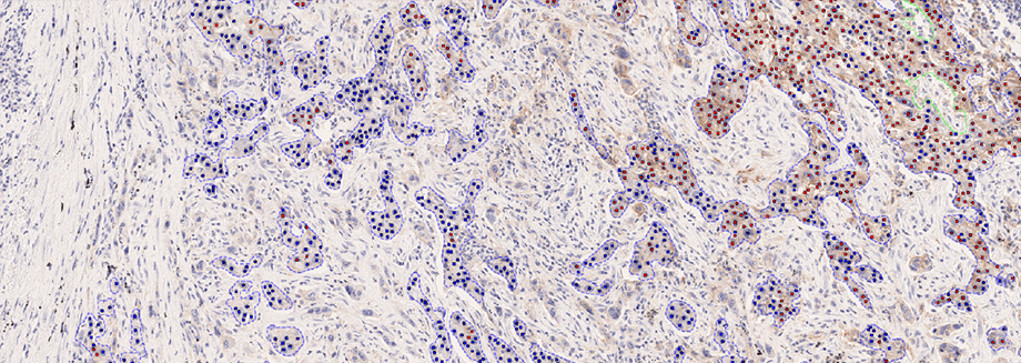
Hørsholm, Denmark — Visiopharm A/S, an innovator in AI-powered digital pathology solutions, has announced the IVDR certification of its PD-L1 APP for non-small cell lung cancer (NSCLC). The APP provides fully automated evaluation for the predictive biomarker, offering whole slide analysis results to the pathologist with no prior interaction needed. With exceptional accuracy and consistency in biomarker scoring related to eligibility for immune checkpoint inhibitors, it integrates seamlessly with PACS/IMS systems used by pathology labs, ensuring optimal workflow efficiency.
The APP is the first IVDR-cleared PD-L1 algorithm in the market and expands Visiopharm’s existing diagnostic portfolio for breast, colorectal, and prostate cancer with lung cancer – the second most common cancer type but with the highest number of deaths per year [1]. The product addresses the needs of the current diagnostic market where prescriptions of certain PD-L1 immune checkpoint inhibitors have grown more than 500% between 2020 and 2021 alone [2]. With a growing technology stack for precisely quantifying predictive biomarkers, it shows Visiopharm’s leading capabilities to support the needs of pharma for precision pathology.
Achieving IVDR requires a significant effort, and the PD-L1 APP is the 9th IVDR clearance for Visiopharm. The validation is based on the results of a comprehensive clinical study encompassing a large patient cohort and evaluations from three distinct European sites. The study directly compared manual assessments with the APP’s stand-alone analysis and APP-assisted interpretations. The results were remarkable, demonstrating substantial to near-perfect agreement with the multi-reader reference score and a high concordance with gold-standard expert manual assessments. Most notably, the data indicates increased agreement rates with pathologists reporting efficiency gains when using the APP, underpinning the fully automated APP’s potential to address the primary challenges of PD-L1 IHC scoring.
Professor Ralf Huss from University Hospital Augsburg, Germany, reflects on the APP’s impact: “After getting acquainted with the APP and building trust in the scores, it was much easier and faster to score PD-L1 NSCLC. It took less than 20 cases doing a site-by-site comparison of the manual scoring with the APP-assisted scores to build up that level of trust. The possibility to interact with the classification and review the image also allows for a very quick plausibility check, if there is some doubt on the APP-generated results, e.g. does the APP only score tumor cells, etc. Now I don’t want to miss the APP anymore to do PD-L1 NSCLC scoring, especially on a larger scale.”
Visiopharm has partnered with Agilent Technologies Inc. to develop and validate the PD-L1 Diagnostic APP adding to its expansive portfolio of fully automated workflow APPs that have been validated using Agilent’s Dako Omnis platform.
Lou Welebob, Vice President and General Manager, Diagnostics and Genomics Group, Agilent comments, “The IVDR certification of Visiopharm’s PD-L1 APP for non-small cell lung cancer (NSCLC) represents a significant advancement in digital pathology. We are excited about the algorithm-assisted advances in pathology scoring and to see how workflow integration may deliver on overall efficiency.”
Dirk Vossen, CDO of Visiopharm, emphasizes the company’s commitment: “Our PD-L1 algorithm exemplifies our drive to refine diagnostic accuracy and support improved efficiencies. Furthermore, it is a product of our fruitful alliance with Agilent.”
Visiopharm® is a leading provider of AI-driven precision pathology software for research and diagnostics. In research, it is a technology leader providing tools that help scientists, pathologists, and image analysis experts produce accurate data for all types of tissue-based research. In diagnostics, it is a leader within clinical applications, with no less than nine diagnostic algorithms cleared under IVDR for EU and UK customers. These applications provide diagnostic decision support and and can be easily activated and integrated into existing lab workflows.
Founded in 2002, Visiopharm is privately owned and operates internationally with over 750 customer accounts in more than 40 countries. The company’s headquarters are located in Denmark’s Medicon Valley, with offices in Sweden, the UK, Germany, the Netherlands, and the United States, and local representation in France and China.
[2] Shin, Y. ., Kumar, A. & Guo, J.J. Spending, Utilization, and Price Trends for Immune Checkpoint Inhibitors in US Medicaid Programs: An Empirical Analysis from 2011 to 2021. Clin Drug Investig43, 289–298 (2023)