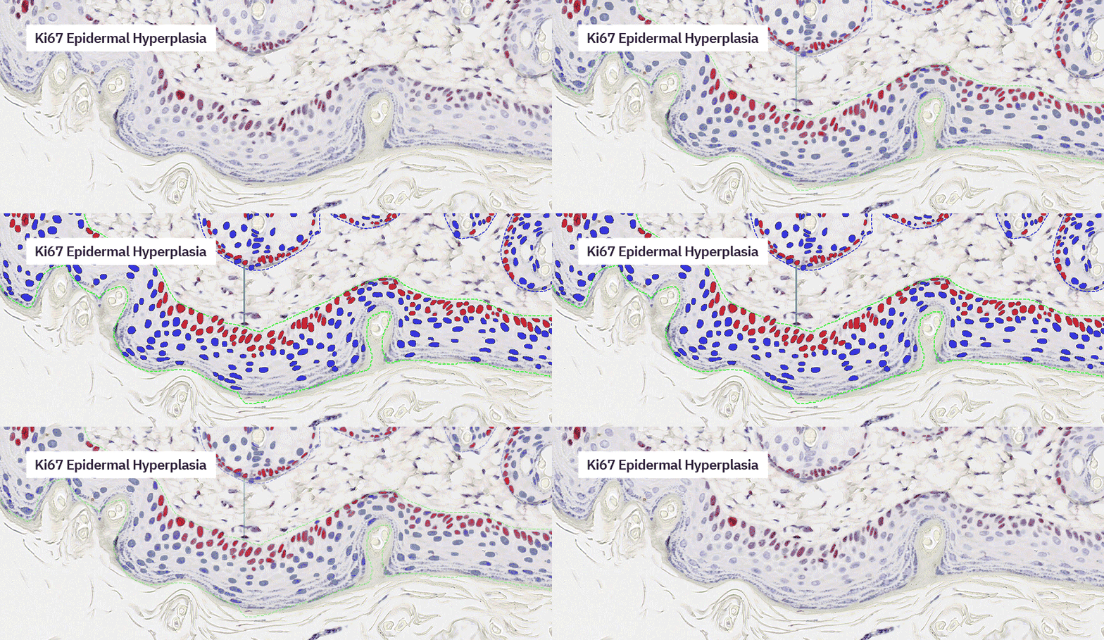
The latest version of our software has arrived and there’s a load of improvements we want to tell you about. We have created a short video to walk you through all the updates – watch it now to see what’s new!
Prefer to read? Keep scrolling to explore the key enhancements.
What’s new
PhenoplexTM Guided Workflow
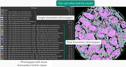
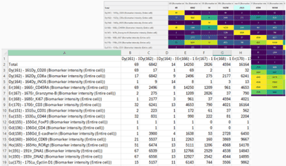
This release includes the possibility to sort the phenotype list by biomarker count and extraction of the co-occurrence matrix data.
It also includes improved loading of data in the Phenoplex Guided Workflow.
Data exploration and QC tool
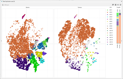
K-means clustering is available for the t-SNE plot and the tool is now dockable like many of the other dialogues and tools in the software.
General
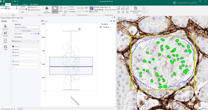
Additionally, the color adjustment is improved for multi-view of images, the heatmap is improved and there are new file formats and layer data that is supported in Visiopharm.
The liver’s microenvironment holds the key to understanding and addressing complex liver diseases, from fibrosis to emerging infections. In this blog, we sit down with Professor Heather Stevenson-Lerner, MD, PhD, a physician-scientist and the sole liver pathologist at the University of Texas Medical Branch (UTMB), whose work spans over a decade of groundbreaking research in liver pathology and immune responses.
Prof. Stevenson-Lerner shares her experience leveraging Visiopharm software to explore the liver microenvironment, offering unique insights into macrophage-mediated immune responses and the use of advanced AI tools for spatial biology analysis.
She also highlights how Visiopharm empowers her team to study liver diseases with unprecedented precision, ensuring reproducible and impactful research outcomes.
Read on for the full interview or watch the video recording to discover how Prof. Stevenson-Lerner is driving innovation in liver pathology.
Visiopharm: Welcome, Prof. Heather, can you tell us a little bit about yourself?
Prof. Heather Stevenson-Lerner: My name is Heather Stevenson-Lerner, I’m a professor in the Department of Pathology at the University of Texas Medical Branch (UTMB) in Galveston, Texas, and a physician-scientist studying the liver microenvironment and various types of chronic liver disease. I’ve been using Visiopharm software for nearly seven years, generating valuable data for our projects. I’m excited to share more about our work today.
Visiopharm: Can you describe the primary focus of your research and the specific pathologies you’re studying with Visiopharm software?
Prof. Heather Stevenson-Lerner: I’ve been the sole liver pathologist at UTMB since 2014, signing out every liver biopsy for over a decade. Visiopharm’s tools allow us to study the hepatic microenvironment using formalin-fixed, paraffin-embedded tissue. With access to a 30-year archive of liver tissue, we’ve analyzed a range of chronic liver diseases, such as steatotic liver disease, viral hepatitis C, autoimmune hepatitis, primary biliary cholangitis, and even infectious diseases like Ebola. At UTMB, we specialize in emerging infections, and the ability to analyze deactivated tissues without requiring high-containment labs has been invaluable.
The primary focus of our research is to study the hepatic microenvironment. We’re very interested in macrophage-mediated immune responses, which play a critical role in promoting inflammation and fibrosis in the liver. We use archived liver biopsies to stain different antibodies, and Visiopharm is integral to our work, especially for multiplex staining. For instance, we use a six-antibody panel for macrophages, and we look at markers in the same compartment on the same cell, and there is no other software platform that allows us to analyse that.
“Over the years, we’ve tried a lot of different things, and we found that Visiopharm is the best at helping us differentiate these different phenotypes within the hepatic microenvironment and these various diseases.”
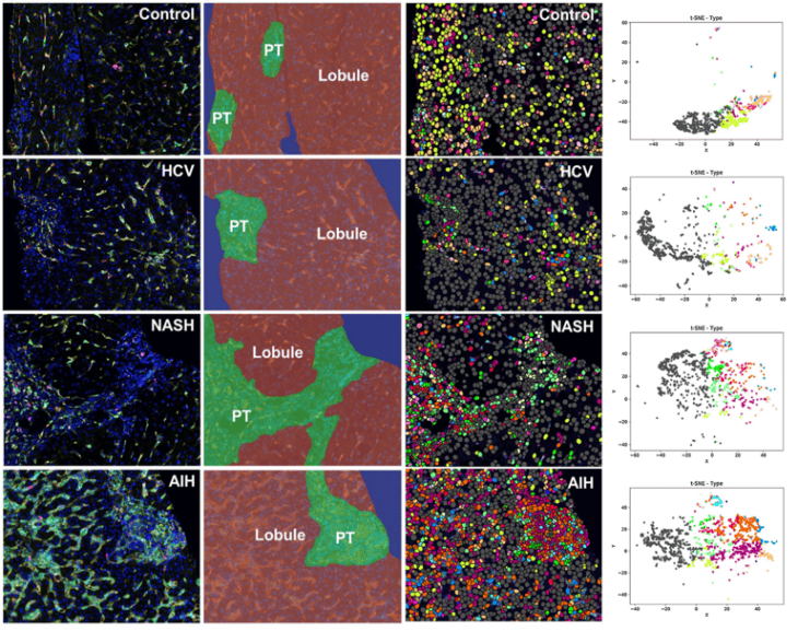
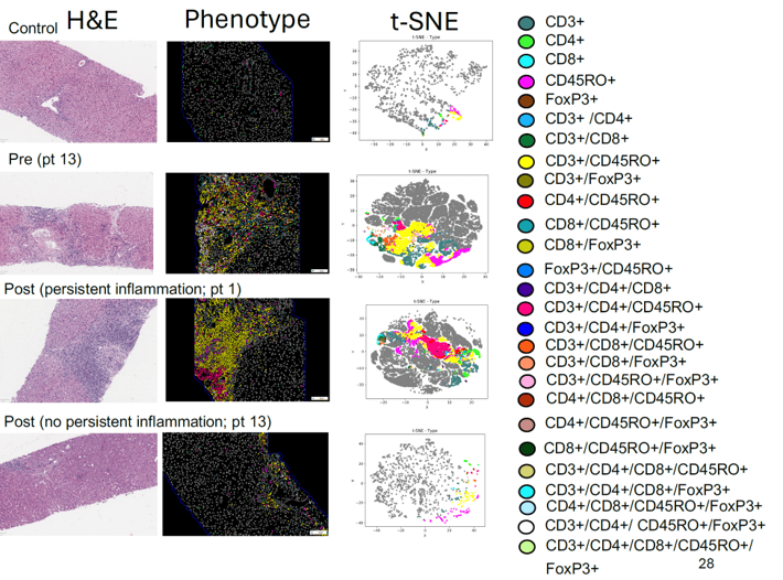
DAPI, CD68 (Kupffer cells), CD14 (pro), CD16 (anti), CD163 (“M2”), Mac387 (Recruited). Tissue segmentation, phenotyping, and data exploration with tSNE plots using Visiopharm software (Phenoplex).
Visiopharm: What challenges or limitations in your research led you to consider using AI solutions like Visiopharm, for example?
Prof. Heather Stevenson-Lerner: I collaborated with Dr. Jared Burks at MD Anderson Cancer Center when we started. Spatial approaches were already well-established for tumor microenvironments, so I thought, why not apply similar techniques to inflammatory liver diseases? We were among the first ones doing it here at UTMB, and now it’s really caught on. Obviously, the spatial field has grown so much in the last few years, but Dr. Jared Burks and I both came to the conclusion that Visiopharm was the best.
“At that time, we tested several different platforms. Each had pros and cons, but Visiopharm stood out, particularly for its AI-driven cell segmentation. Accurate cell segmentation at the initial step is crucial since it ensures precise downstream data analysis.”
In addition, when we first started using Visiopharm, we wanted to make sure that the data was reproducible. You know, patients are different. We have to make sure what we’re seeing is accurate. To validate our results, we collaborated with the bioinformatics group at the University of Michigan, which is one of the best in the country. We compared results from the same experiments using two different analysis methods: multicomponent TIFF files processed with Visiopharm and numerical cell segmentation data from InForm software (Vectra Polaris). And we did come up with the same. We have the same results by looking at the data in two completely different ways. The advantage of Visiopharm is that we can perform the analysis independently in our own lab without relying on external support.
Visiopharm: How has the use of Visiopharm software improved the quality and accuracy of your research outcomes?
Prof. Heather Stevenson-Lerner: We’re really trying to preserve the spatial aspects and keep the liver architecture intact. There are many great things out there. For example, when I was a graduate student, I did flow cytometry in the liver. But with flow cytometry, you have to homogenize the entire liver tissue. First, you need fresh tissue—not formalin-fixed—which can be challenging, especially with human patients. When you homogenize the liver, you separate the cells but can’t determine their spatial location. For example, is a cell in the portal tract or the lobule of the liver? What types of cells are next to each other? Who is interacting with whom? You can’t perform this kind of neighbor analysis with flow cytometry.
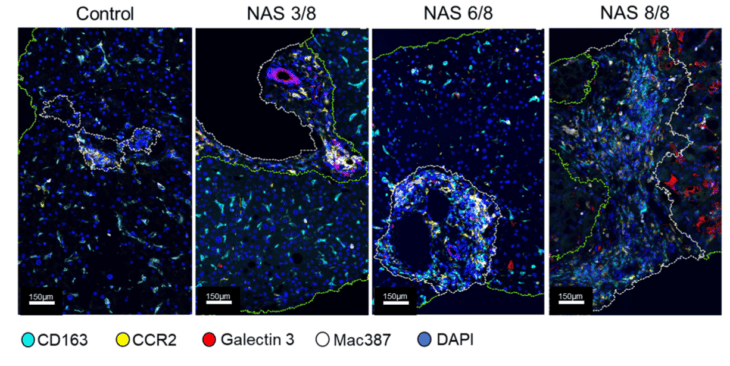
Analysis of individual patients for variations in druggable targets (CCR2 and Galectin 3). Representative images with tissue segmentation into portal tract and lobular regions using Visiopharm software for image analysis (Phenoplex).
Similarly, single-cell RNA sequencing recreates the liver microenvironment but still doesn’t preserve the architecture. Visiopharm allows you to analyze different cell types while maintaining spatial relationships. You can identify phenotypes, measure marker expression levels, and perform tSNE to study the relationships between cells. This ability to preserve liver architecture is something that the Visiopharm analysis enables that other applications cannot. Additionally, when looking for small signals, we also use NanoString to analyze whole RNA signatures from homogenized liver tissue. While NanoString is powerful, homogenization can dilute specific signals, such as those from a single cell type with a druggable target or marker expression. With Visiopharm, I can go directly into the liver tissue, segment CD68-positive macrophages, cluster them, and analyze their signals. This allows me to identify data I might miss when homogenizing the entire liver, making Visiopharm an incredibly powerful tool.
Liver biopsies from patients with HCV analyzed with Visiopharm software (Phenoplex).
Visiopharm: In which ways did Visiopharm software prove to be the only viable solution for your research needs compared to other tools or methods you considered?
Prof. Heather Stevenson-Lerner: Well, I think there are several important things. As I mentioned, being able to stain multiple antibodies in the same cellular compartment is crucial. We try to combine different types of antibodies—some that are nuclear, cytoplasmic, or membranous—which helps. However, we also work with antibodies like CD68, CD163, CD14, and CD16, which are all in the same compartment but labeled with different fluorophores or Opal fluorophores. Visiopharm has been the best at accurately separating these cells with antibodies in the same compartment.
Another key advantage is the ability to use pre-existing algorithms from Visiopharm’s website. Once we develop customized algorithms, we can apply them to different chronic liver diseases with minimal adjustments. For example, we created an algorithm for chronic hepatitis C (HCV) and, with slight modifications, applied it to patients with autoimmune hepatitis. This flexibility is especially helpful when using the same macrophage antibody panel across various diseases.
Additionally, the training sessions with Visiopharm have been invaluable. These sessions are included in our software package, and we’ve had weekly meetings during heavy usage periods or when addressing software updates or specific questions. This ongoing training has been crucial to our success.
“Finally, Visiopharm’s customer support has been excellent. They are always available to assist whenever we have questions, ensuring we’re never held up in our research. These aspects—antibody differentiation, customizable algorithms, training, and support—are the main things that have been extremely helpful for us.”
Are you curious to see the work of Prof. Heather Stevenson-Lerner? Check out these recent publications:
Hørsholm, Denmark — Visiopharm, a world leader in AI-driven precision pathology, is thrilled to become a preferred partner to PathAI with the integration of its precision pathology APPs into the AISightⓇ1 Image Management System (IMS). This partnership underscores Visiopharm’s commitment to transforming diagnostic and translational pathology workflows by enabling PathAI’s AISight IMS users to analyze images with unparalleled accuracy and speed.
Visiopharm offers a comprehensive suite of diagnostic APPs, including a breast panel (HER2, ER, PR, metastasis detection, and Ki-67), and a non-small cell lung cancer (PD-L1) APP. These APPs are IVDR-cleared for use in the EU and UK and designated for research use in other regions. These tools will empower pathology laboratories with high-performance algorithms designed to streamline workflows and enhance diagnostic precision.
PathAI’s AISight IMS is designed to enhance interoperability and efficiency in digital pathology. Laboratories using AISight can seamlessly access a diverse portfolio of digital pathology algorithms from leading global AI providers, including Visiopharm. Pathologists will benefit from rich visual support, including AI-generated overlays and embedded quantitative results, driving more informed decision-making and differentiated workflows.
“We are excited to collaborate with Visiopharm and integrate their leading AI image analysis technologies into our AISight platform,” said Andy Beck, CEO of PathAI. “This partnership reinforces our mission to provide pathologists with comprehensive, state-of-the-art tools that enhance precision and increase workflow efficiency in the pathology laboratory. By incorporating Visiopharm’s innovative solutions, we are broadening access to top-tier AI capabilities, ultimately driving better patient outcomes and setting a new standard in digital pathology.”
Michael Grunkin, CEO of Visiopharm, expressed his enthusiasm about the partnership: “We are delighted to collaborate with PathAI to bring our portfolio of fully automated AI-driven diagnostic and translational solutions for breast and lung cancer to a broader audience. Our commitment is to expand access to precision pathology on leading platforms. Becoming a preferred partner for PathAI’s AISight Image Management System is an important step towards this goal. With our platform-agnostic technology designed for seamless integration, we look forward to working closely with the PathAI team to accelerate the adoption of digital pathology worldwide, ultimately enhancing lab efficiency and improving the quality of patient care.”
Pathology laboratories worldwide are invited to experience the power of Visiopharm’s precision pathology APPs on PathAI’s AISight IMS. To schedule a demonstration of the APPs, please contact us at https://visiopharm.com.upd.mtra.in/.
1AISight is for Research Use Only in the US; AISight Dx is CE-IVDR in Europe and UKCA in UK
About Visiopharm
Visiopharm is a leading provider of AI-driven precision pathology software for research and diagnostics. In research, it is a technology leader providing tools that help scientists, pathologists, and image analysis experts produce accurate data for all types of tissue-based research. In diagnostics, it is a leader within clinical applications, with no fewer than nine diagnostic algorithms cleared under IVDR for EU and UK customers. These applications provide diagnostic decision support and can be easily activated and integrated into existing lab workflows. Founded in 2002, Visiopharm is privately owned and operates internationally with over 750 customer accounts in more than 40 countries. The company’s headquarters are located in Denmark’s Medicon Valley, with legal entities in Sweden, the UK, Germany, the Netherlands, and the United States, and local representation in France and China.
About PathAI PathAI is a global leader in AI-powered digital pathology solutions, dedicated to improving operational efficiency in pathology labs worldwide. Through its innovative technology and strategic partnerships, PathAI is shaping the future of medical diagnostics and advancing patient care across the globe.
Los Angeles, CA – Molecular Instruments® (MI), the inventor of the HCR™ Imaging technology, is pleased to announce a strategic partnership with Visiopharm, a global leader in AI-driven precision pathology software. This collaboration brings together MI’s clinical-grade HCR™ platform with Visiopharm’s Oncotopix® Discovery software, empowering users with a highly flexible, deep learning approach to precise and efficient image analysis for RNA-ISH assays.
MI’s class-leading HCR™ Pro RNA-ISH assay, lauded for making protease-free sample preparation the standard for RNA-ISH, provides unparalleled specificity and sensitivity in RNA detection and enables seamless integration with existing IHC/IF workflows. When combined with Oncotopix® Discovery, researchers can analyze chromogenic and fluorescent HCR™ Pro RNA-ISH and HCR™ Pro RNA-ISH + IHC/IF co-detection images with more precise segmentation and spot counting, ensuring accurate gene expression profiling in even the most challenging samples.
“Our North Star at MI is to set bioimaging assays to a true ‘clinical-grade’ standard, focusing on performance, robustness, and accessibility,” said Dr. Aneesh Acharya, Chief Commercial Officer at Molecular Instruments. “Partnering with Visiopharm reflects our shared drive to delivering clinical-grade assays, paired with world-class analysis tools that empower pathologists and support decision-making. We’re thrilled to kickstart this journey with the team at Visiopharm to bring new possibilities to RNA-ISH.”
Visiopharm’s Oncotopix® Discovery software enables users to explore complex tissue data intuitively and customize analyses to fit their specific experimental needs. Its advanced exploratory capabilities enhance analysis accuracy in complex tissue samples. The ability to develop custom deep-learning-based segmentation algorithms, which can account for both cellular and nuclear morphology, enables more precise quantification of RNA. This marks a significant improvement over traditional methods, which often struggle with complex or irregular structures, such as anuclear or multinucleate cells, leading to missed or inaccurate counts.
“At Visiopharm, our mission is to empower researchers with advanced tools that deliver unmatched precision in image analysis,” said Regan Baird, Senior Vice President, Commercial Strategy Deployment of Visiopharm. “Partnering with Molecular Instruments allows us to elevate the standard of RNA-ISH analysis, integrating our Oncotopix® Discovery software with MI’s HCR™ technology to provide researchers with an unprecedented level of accuracy and efficiency. We are excited to collaborate and advance the field of precision pathology, accelerating breakthroughs in disease research and diagnostics.”
This partnership marks a pivotal advancement in research, combining high-performance, clinical-grade RNA-ISH with sophisticated image analysis. Together, we are equipping scientists to drive breakthroughs in disease studies, offering deeper insights into gene expression and protein interactions. By integrating our innovative technologies, we aim to accelerate discoveries across translational medicine, where accurately and precisely understanding gene and protein expression are essential.
About Visiopharm:
Visiopharm is a leading provider of AI-driven precision pathology software for research and diagnostics. In research, it is a technology leader providing tools that help scientists, pathologists, and image analysis experts produce accurate data for all types of tissue-based research. In diagnostics, it is a leader within clinical applications, with no fewer than nine diagnostic algorithms cleared under IVDR for EU and UK customers. These applications provide diagnostic decision support and can be easily activated and integrated into existing lab workflows. Founded in 2002, Visiopharm is privately owned and operates internationally with over 750 customer accounts in more than 40 countries. The company’s headquarters are located in Denmark’s Medicon Valley, with legal entities in Sweden, the UK, Germany, the Netherlands, and the United States, and local representation in France and China.
Molecular Instruments® (www.hcrimaging.com) develops and synthesizes molecular kits powered by the breakthrough HCR™ imaging platform for applications in academic research, drug development, synthetic biology, and clinical pathology and diagnostics.
Welcome to our latest blog interview, where we spotlight Glint Lab, a full service histopathology and digital pathology lab located in San Diego, California. Today, we’re joined by three of their leading experts—Chief Scientific Officer Oanh Nguyen, Director of Image Analysis Jason Roberts, and Chief Executive Officer Misagh Naderi, to explore how they’re leveraging Visiopharm’s advanced image analysis software to address some of the most complex scientific questions.
In this insightful conversation, they share their experience implementing Visiopharm software, highlight key features that are driving their research forward, and showcase innovative ways they are using digital pathology to unlock new insights in the field.
Oanh Nguyen Chief Scientific Officer at Glint Lab I am a scientist with a strong background in histopathology and assay development/optimization for advanced tissue-based assays, including IHC or mIHC, immunofluorescence or mIF, and in situ hybridization, along with other advanced protein-RNA multiplex assays. My expertise lies in enhancing these assays to obtain actionable quantitative data and insights for scientific research. With experience working in both clinical diagnostic lab environments and the preclinical and discovery research space, I bring an understanding of both clinical and research-based applications and the needs of our clients, helping to design and develop assays from the earliest stages for potential clinical applications.
Jason Roberts Director of Image Analysis at Glint Lab
I am a data scientist dedicated to advancing computational pathology and spatial biology through innovative machine learning solutions. Drawing on my deep experience in image analysis, I engineer computer vision algorithms and leverage statistical models to transform biological data into meaningful insights. Through my work, I strive to bridge the gap between data science techniques and biological research, ultimately contributing to more effective diagnostic tools and therapeutic strategies.
Misagh Naderi Chief Executive Officer at Glint Lab I began my career as an engineer before transitioning into biology, building a portfolio of research publications in molecular virology and computational biology. My experience extends to technology commercialization and IP law, particularly in life science technologies that integrate AI and machine learning. For the past three years, I’ve had the privilege of working on projects that blend technology, histology, and biology in innovative ways. As CEO of Glint Lab, I’m fortunate to collaborate with a team of experts focused on leveraging technology across the workflow and in image analysis to enhance quality and produce quantitative, actionable insights for research and clinical trials. My goal is to keep learning and contributing to advancements that lead to better treatments and improved patient outcomes.
Can you tell us a bit about Glint Lab and which services you are offering to your customers?
Misagh Naderi: Glint Lab is a full service histopathology and digital pathology laboratory based in San Diego, California. We are a team of seasoned lab and data scientists dedicated to revolutionizing preclinical and early drug discovery through our comprehensive suite of precision pathology services.
We pride ourselves on delivering high quality tissue processing, sectioning, histology, and cutting-edge image analysis services.
Unlike traditional labs, we view our relationship with clients as a partnership. We go beyond transactional services to foster meaningful collaborations that drive scientific discovery. For example, our clients can consult with us prior to project initiation in order to prepare the study design and the budget that relates to their histology and data analysis needs. Our clients’ success is our success, and we are committed to delivering the best data possible to fuel their research endeavors.
Since when have you been using Visiopharm and what led you to Visiopharm originally?
Jason Roberts: Visiopharm was among the first systems we integrated at the launch of Glint Lab earlier this year, but our team has extensive experience with the software. Visiopharm offers us the unparalleled ability to develop robust custom algorithms for a wide variety of image analysis applications, and to explore spatial data in ways that can not be done with comparable tools. In the rapidly evolving field of digital pathology, integrating Visiopharm’s AI-driven image analysis software is transforming our approach to understanding complex biological systems.
“Visiopharm offers us the unparalleled ability to develop robust custom algorithms for a wide variety of image analysis applications, and to explore spatial data in ways that can not be done with comparable tools.”
Which features of Visiopharm do you consider most impactful thus far?
Jason Roberts: We greatly value the platform’s cutting edge deep learning classifiers and neural network architectures which we employ for a multitude of image processing tasks. The professional and enhanced modules enable us to perform image preprocessing, automated array handling, image coregistration, and neural network parameter optimization for our most complex and challenging studies. The interactive data exploration tool is one of our favorite features and certainly sets Visiopharm apart. It empowers us to generate captivating, feature-rich visualizations to examine cellular relationships in context. This approach results in the deepest possible insights for our client’s tissue data – effectively driving informed decisions and propelling their research forward.
“The interactive data exploration tool is one of our favorite features and certainly sets Visiopharm apart.”
Phenoplex in action: Glint Lab harnesses advanced data exploration tools for precise navigation and insights.
We also believe that cooperation and long term partnerships are fundamentally important to being successful in the dynamic biotechnology research industry. Visiopharm’s collaborative ethos and commitment to innovation in digital and computational pathology made choosing their tools a clear and easy decision. We appreciate the opportunity to work with the Visiopharm team, and look forward to testing out new features, providing feedback, and playing an active role in the continued development of their image analysis software.
Can you describe the process of implementing Visiopharm and how your experience has been working with our team?
Jason Roberts: Our experience with the Visiopharm team has been exceptional. We are very grateful to work with such an experienced, professional, and knowledgeable group of people. The folks we’ve worked with across professional services, sales, marketing, leadership, and engineering have been instrumental in implementing our image analysis services.
Jeni Caldara, Strategic Partnerships Manager, has been a considerable asset to our organization and has offered a plethora of strategies to both maximize and streamline our use of the software to best serve our clients needs. Brenna O’Neill, Application Support Specialist, has provided us with incredibly valuable training and onboarding sessions, and is always quick to respond with creative and effective solutions to our technical questions. Raymond Brandenburg, IT Solutions Architect, has supported us in the configuration of our powerful cloud computing environments, and advised our team on inventive IT integration practices. We look forward to expanding our relationship with the Visiopharm team as our organization continues to grow.
Which kinds of problems or questions of your customers can you address with Visiopharm?
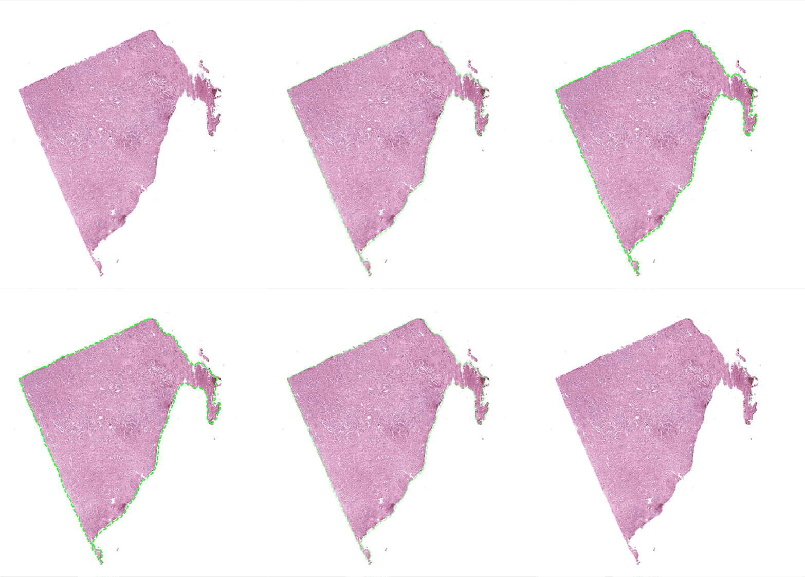
Prostate – Tissue detection
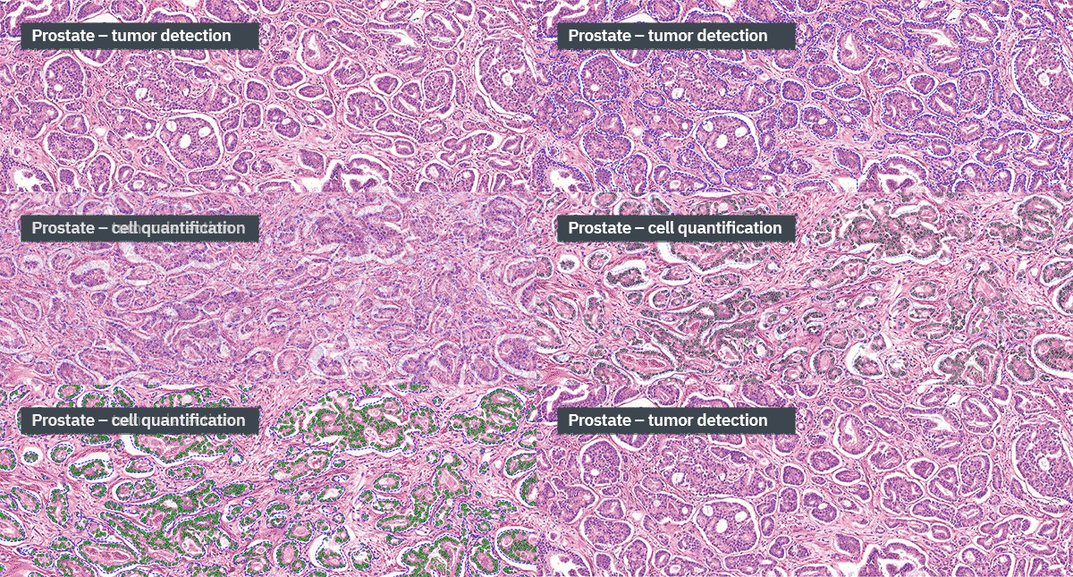
Oanh Nguyen: At Glint Lab, we use Visiopharm’s advanced image analysis software to tackle a wide range of challenges faced by our clients. From IHC/IF or mIHC/mIF to special stains to RNA ISH, we provide standard image analysis quantitative outputs and custom solutions. With Visiopharm, we excel in spatially resolved quantification, enabling us to perform complex analyses such as clustering, neighborhood mapping, proximity assessments, and evaluations of aggregation, infiltration, density, and dispersion. Our expertise spans multiple disease areas, including Toxicology (e.g., toxicity in rodent tissues), Oncology (e.g., tumor microenvironment analysis in human samples), Immunology (e.g., immune cell profiling in human, rodent and other tissues), Musculoskeletal disorders (e.g., Laminin/Dystrophin analysis in Duchenne Muscular Dystrophy (DMD) models, centralized nuclei analysis in muscular dystrophy X-linked mouse (mdx) models), Cardiometabolic diseases (e.g., adipose tissue analysis in pig models), Nephrology (e.g., glomerular assessments in human and rodent kidney tissues), NASH (e.g., fibrosis evaluation in rodent liver models), and more. We also have extensive expertise across various species and tissue types, including Rodents, NHP (Non-Human Primates), Pigs, Rabbits, Humans, Organoids, and others, ensuring that your data is examined with the most relevant biological context. Visiopharm’s APP center algorithms further enhance our capabilities, allowing for custom analysis and visualization that meet the specific needs of your project. Through this partnership, Glint Lab and Visiopharm empower you to gain deeper insights from your tissue samples, providing the robust, accurate, and actionable data needed to advance your research.
“With Visiopharm, we excel in spatially resolved quantification, enabling us to perform complex analyses such as clustering, neighborhood mapping, proximity assessments, and evaluations of aggregation, infiltration, density, and dispersion.”
Can you share some of the creative ways you use our software?
Jason Roberts: We deployed VIS in a powerful cloud environment which optimizes compute and storage while maintaining maximum usability for both our data scientists performing image analysis and our clients. Taking advantage of our unique cloud architecture, we’ve developed an advanced data analytics platform to be used by our clients and collaborators. Here’s how it works: we share a secure and easy-to-access VIS viewer with researchers who want to review their results and carry out granular downstream analysis using Visiopharm’s data exploration tools. Our advanced analytics platform powered by Visiopharm allows our clients, whether they are scientists, pathologists, or bioinformaticians, to efficiently review and investigate their data, elucidate spatial cellular relationships, and generate publication-ready visualizations.
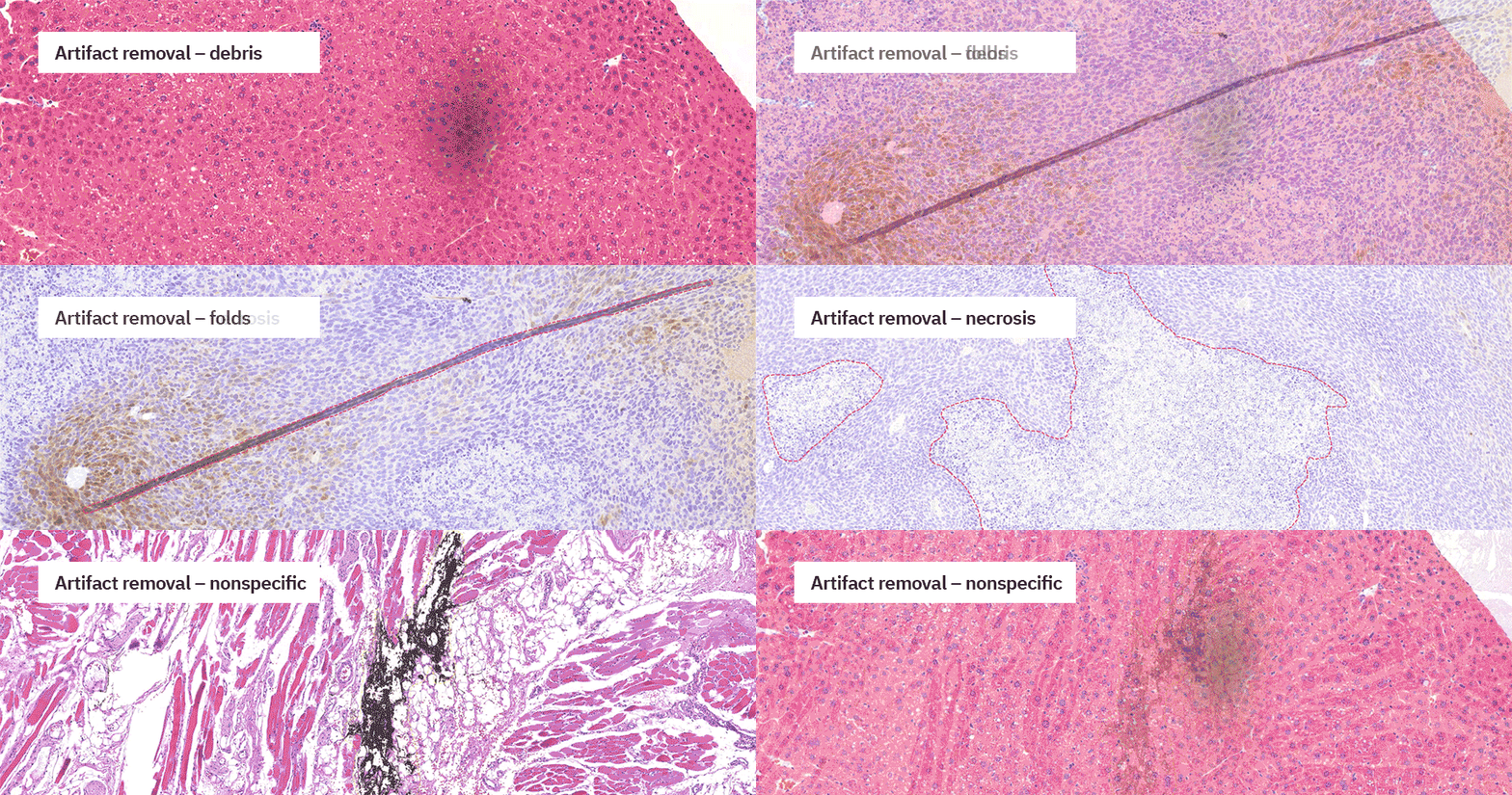
We also leverage Visiopharm to engineer robust quality control and staining consistency assessment algorithms. Our team has developed APPs that automatically identify and exclude pre-analytical artifacts including: folds, blur, slide debris, out of focus areas, nonspecific staining, blood, and more. In addition, we’ve built tools to assess staining quality and consistency of H&E, IHC, and IF images through detailed evaluation of staining intensities across images or batches in a given project. These APPs are always deployed at the beginning of a study, and they are essential in maintaining our rigorous quality standards at Glint Lab.
Robust quality control and staining consistency tools with Visiopharm software.
We benefit from Visiopharm’s bidirectional integrations with other digital pathology software as well, including our image management system. We engage the integration to easily import newly-scanned samples into VIS with only a few clicks, as well as pathologist’s annotations for use as analysis ROIs or as input data for training new models.
“Visiopharm’s valuable tools and integrations drive substantial increases in productivity and enhance our overall customer experience.”
Can you describe the advantages of using your data analysis platform powered by Visiopharm?
Misagh Naderi: After analyzing the images and providing quantitative results, we share an easily accessible instance of Visiopharm with our clients. Here, they can further explore cutting edge algorithms such as clustering methods to dig deeper into the quantitative data that our expert image analysis team has extracted for them. We believe that providing high quality data together with Visiopharm’s tools to transform the data will allow our clients, who are experts in their respective fields, to come up with the best informed hypotheses for future studies. Again, our clients’ success is our success, and we extend all of our resources to make sure they get the most value out of their projects with us.
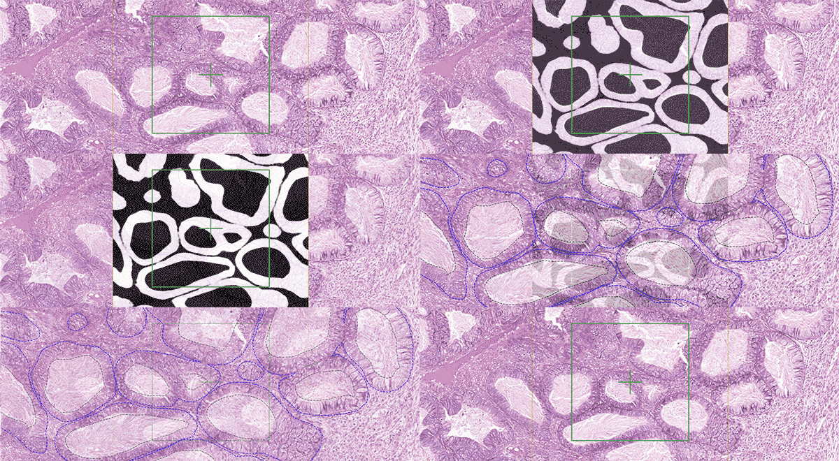
Ovarian tissue analysis and neural network probability feature map
Can you give examples of exciting future applications for Visiopharm at Glint Lab?
Jason Roberts: We look forward to implementing Visiopharm to analyze more multiplex IHC and multiplex IF assays in order to dissect and examine intricate cellular interactions, phenotypes, and biomarker distributions. We also look forward to using Visiopharm’s comprehensive tools to interpret spatial transcriptomics data, allowing us to map gene expression patterns within their native tissue architecture and advance our knowledge in spatial biology. Next-generation neighborhood analysis tools further enable us to investigate microenvironmental interactions at a cellular level, revealing how cellular neighborhoods influence disease progression. Visiopharm’s image analysis solutions are transforming digital pathology, offering profound improvements in patient care, and paving the way for an exciting and optimistic future of personalized medicine. At Glint Lab, we are honored to be a part of this journey.
Wherever your research takes you, we will be there
Glint Lab’s success story is a testament to the power of integrating cutting-edge technology with scientific expertise to tackle challenging research questions. By combining Visiopharm’s powerful image analysis tools with their deep understanding of pathology and data science, they’re pushing the boundaries of what’s possible in preclinical and drug discovery research.
If you have any questions or would like to learn more about their work, feel free to reach out to the Glint Lab team directly at scientist@glintlab.com
To explore how Visiopharm can support your own research, contact us to learn more about our solutions and services.
Hørsholm, Denmark — The University Medical Center Groningen (UMCG) based in the Netherlands is pleased to announce a significant strategic partnership with Visiopharm, a leading global provider of digital pathology solutions, for the coming five years. This collaboration represents an exciting step towards advancing the clinical implementation of AI-driven Precision Pathology.
The partnership revolves around multiple areas of cooperation, including the introduction of a state-of-the-art, automated, AI-based image analysis workflow for digital pathology built upon Visiopharm’s Oncotopix® and Qualitopix® platforms. This comprehensive workflow includes automatic slide quality assessment, continuous stain quality assessment, and biomarker quantification with the overarching objective of enhancing precision within laboratory operations.
The UMCG as a reference center for IHC Precision Pathology
A key initiative of this partnership is the establishment of the UMCG as a reference center. This center is not just a hub of technological innovation but also a living showcase demonstrating the significant impacts of AI-driven precision pathology on the standardization of diagnostic processes and the subsequent elevation of patient care standards. The UMCG is committed to not only adopting these advanced practices but also to meticulously quantifying their benefits and sharing these insights with the global clinical pathology community. The partnership with Visiopharm is in complete alignment with UMCG’s objective of data-driven research, data quality, and, ultimately, improved patient outcomes.
“By joining forces with Visiopharm, we are set to demonstrate the concrete benefits and transformative power of AI in precision pathology,” said Dr. Bert van der Vegt, Clinical Pathologist and lead of the Digital Computational Pathology Unit at UMCG. “As a reference center, we’re excited about the opportunity to share our journey and experiences, showcasing the tangible improvements in standardization and patient care that this technology brings.”
Visiopharm, a trusted name in the field of digital pathology, is equally enthusiastic about this partnership. “UMCG’s dedication to advancing precision pathology, and thereby optimal treatment decisions, perfectly aligns with our corporate mission. We look forward to our partnership and the opportunity to further document the benefits for diagnostic pathology laboratories seeking to implement precision pathology,” commented Dirk Vossen, Chief Diagnostic Officer at Visiopharm.
For clinical pathology laboratories considering the adoption of digital pathology, this private-partnership represents the epitome of innovation and progress. “UMCG and Visiopharm are working collaboratively to usher in transformative changes by offering enhanced data accuracy, operational efficiency, and improved patient care” adds Jacko Duker, project leader Digital and Computational Pathology at UMCG.
For further details about UMCG’s and Visiopharm’s strategic partnership, please visit
About Visiopharm
Visiopharm is a leading provider of AI-driven precision pathology software for research and diagnostics. In research, it is a technology leader providing tools that help scientists, pathologists, and image analysis experts produce accurate data for all types of tissue-based research. In diagnostics, it is a leader within clinical applications, with no fewer than nine diagnostic algorithms cleared under IVDR for EU and UK customers. These applications provide diagnostic decision support and can be easily activated and integrated into existing lab workflows. Founded in 2002, Visiopharm is privately owned and operates internationally with over 750 customer accounts in more than 40 countries. The company’s headquarters are located in Denmark’s Medicon Valley, with legal entities in Sweden, the UK, Germany, the Netherlands, and the United States, and local representation in France and China.
The University Medical Center Groningen (UMCG) is one of the largest hospitals in the Netherlands. More than 12,000 employees work together on care, research, training, and education with the common goal of building the future of health. The UMCG offers complex, academic hospital care. It strives to do this better every day, constantly pushing the boundaries of diagnostics and treatments. The UMCG makes use of knowledge, research, and innovative solutions, as well as offering future-oriented education and training for healthcare professionals. Long-term partnerships build a healthcare landscape that is ready for the future.
The UMCG researchers are pioneers in research who work on relevant research topics and are provided with a broad range of top-notch research facilities. The groundbreaking work of leading national and international researchers makes the UMCG a pioneering center for medical research.
Role of the UMCG Innovation Center
The UMCG Innovation Center has been instrumental in setting up the partnership, facilitated the work on innovative pathology solutions, and continues to support the collaboration.