Author: Visiopharm

  • Straticyte™ – A digitally delivered oral cancer predictor – powered by Visiopharm analysis 

    Straticyte™ – A digitally delivered oral cancer predictor – powered by Visiopharm analysis 

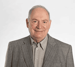
    Kenneth P.H. Pritzker, M.D., BSc (Med), FRCPC

    Kenneth P.H. Pritzker
    M.D., BSc (Med), FRCPC

    Dr. Ken Pritzker served as Chief, Pathology and Laboratory Medicine, Mount Sinai Hospital, Toronto from 1986 to 2008 and as a founder of Mount Sinai Services, a laboratory service outreach vehicle for the biomedical research and innovation sector.

    With over 290 scientific publications and 27 book chapters, Ken is a recognized leader internationally in the fields of arthritis, biomaterials, genomics, cancer diagnostics and nanotechnology. Throughout his career, Ken has served in leadership and advisory positions to national and international professional societies, research institutes and healthcare organizations.

    Ken Pritzker co-founded the company Proteocyte AI in 2011. The company just brought to market their first prognostic LDT test for oral pre-cancer lesions, Straticyte.

    Visiopharm: Can you tell us more about your company Proteocyte and how it all began?

    Dr. Ken Pritzker: Sure, it all started several years ago, when Dr. Ranju Ralhan and Michael Siu discovered, that the nuclear accumulation of S100A7 could serve as a predictor of poorer prognosis for Head and Neck squamous cell carcinoma and could predict a higher risk of transformation of oral premalignant lesions.1 We founded Proteocyte to dive deeper into this promising prognostic biomarker. Even though this finding could be applied to different cancer types, oral cancer was of particular interest to us, due to its high mortality of 50%, which is largely related to late diagnosis. Those late-stage lesions are very costly and difficult to treat. A high percentage of these cancers develop over multiple years in patients who have atypia initially, so they get a biopsy, but it often does not reveal pathologic dysplasia in the histopathologic examinations. Also, the histopathological dysplasia grading, which shows high inter- and intraobserver variation, does not relate very well to the risk of developing cancer from the lesion.2,3 However, some of those samples with mild dysplasia do show the S100A7 accumulation and thus have an elevated Straticyte value. So this is a very important finding because it alerts the clinicians to follow the patient fairly closely, despite their low histopathological grading.
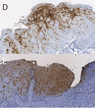
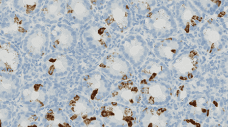
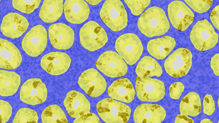
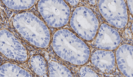
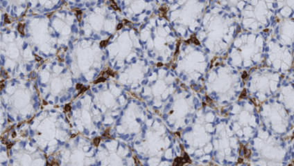
    Immunohistochemistry staining of S100A7 biomarker

    Figure 1 Immunohistochemistry staining of S100A7 biomarker in D) STRATICYTE Medium Risk E) STRATICYTE High Risk; From: BM Renick and MR Darling, Clinical Management of Patients With Oral Epithelial Dysplasia And Elevated STRATICYTETM Risk For Progression To Cancer, Poster @ IAOO, 2019

    Visiopharm: What led you to choose Visiopharm and how has it been working with the company?

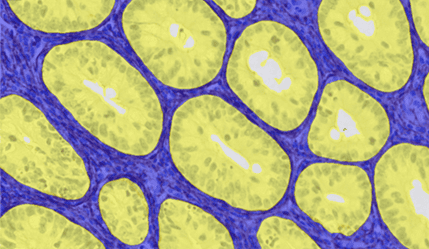
    Dr. Ken Pritzker: We already knew the company and had worked with the software for several years back then at the Mount Sinai Hospital in Toronto. Already at this time, Visiopharm was one of the market leaders of morphometry applied to digital images of tissue slides. We already had a good and long relationship with them and the team, who provided advice. Back then it was just a few people, and we had a great relationship. They have been excellent, and the quality of support has been really superb, so we’ve been very happy with it. Visiopharm software is used to quantitate the immunohistochemical marker and it’s also used to measure certain morphological aspects of the cells. With the support of the Visiopharm team, we have created several APPs in Visiopharm, covering the whole workflow from region detection to score calculation.

    Visiopharm: So how does Straticyte work and how can physicians use it?

    Dr. Ken Pritzker: Our product Straticyte is an LDT test and it combines the evaluation of this specific IHC cell marker with an evaluation of cell morphometry. There are very few, if any, other biomarkers products out there which have a combination of two very different modalities. So this was an innovation. The other is that we saw ourselves from the beginning as an information company. The idea would be that the image of the oral epithelium would be transmitted to the company for analysis, and that we would have a serial database from which we could continue to develop the product and that has been a successful approach. Straticyte can calculate the 5-year probability that dysplastic lesions will progress to cancer. It has a sensitivity of 96% and a NPV of 95%, and those are significantly higher than the values for histological dysplasia grading, which are 75% and 59% respectively.4 The test is so good that if the Straticyte value is low, the patient can go back to live a normal life. But for cases with an elevated Straticyte value, those patients need to be watched closely and followed up against a possible recurrence or cancer. So compared to the current practice, Straticyte provides greater objectivity, sensitivity, and predictive power.

    The way it works is that the physicians anywhere take a biopsy and that is assessed by the pathologist for whether there is pre-cancer present. If so, the pathology lab forwards unstained slides to one of our reference laboratories, which are contracted with Proteocyte and this lab performs the stains. The scanned image is then sent to us and we analyze the image and issue a report.
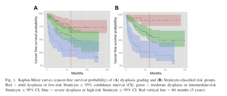
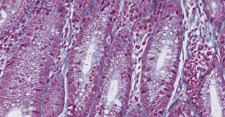
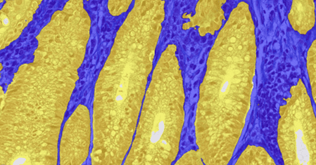
    Figure 2: From: Hwang JTK et al, 2017, doi: 10.1016/j.oooo.2016.11.004

    Visiopharm: How did you develop and validate your product?

    Dr. Ken Pritzker: We started with samples from 250 patients with oral pre-cancer. From there we expanded to other sites in Canada, a site in Ireland and several sites now in the United States. By now, we have a database of more than 600 patients with clinical follow up. That’s probably the largest database of oral pre-cancer that exists, because typically, these patients present to local hospitals and if they have pre-cancer, they may not be followed up very well. So we know exactly the relationship between the test and the prognosis out beyond five years. It’s now more than ten sites and very different populations. And we noticed that lesions from different places have different characteristics. Those might reflect the state of health care in different places or the state of nutrition and this cannot be picked up if you do a single-site database.  

    At this point, the algorithm is very mature and our laboratory has received ISO15189 and CLIA accreditation. In the background we work on improving the tests, but it requires a considerable validation operation to move it from the version which we’re using, which is quite good to the next version, which might be a bit better.

    Learn more about Straticyte.

    ——————————————————————————————————————–

    Main references related to the interview
    1. Tripathi et al, 2010, DOI: 10.1371/journal.pone.0011939 ↩︎
    2. Dost F, Le Cao K, Ford PJ, Ades C, Farah CS. Malignant transformation of oral epithelial dysplasia: a real-world evaluation of histopathologic grading. Oral Surg Oral Med Oral Pathol Oral Radiol. 2014;117:343-352. ↩︎
    3. Fleskens S, Slootweg P. Grading systems in head and neck dysplasia: their prognostic value, weaknesses and utility. Head Neck Oncol. 2009;1:11. ↩︎
    4. Hwang et al, 2017, DOI: 10.1016/j.oooo.2016.11.004 ↩︎
  • Visiopharm announces collaboration with Glint Lab, Inc to enhance pathology solutions and drive scientific discovery

    Visiopharm announces collaboration with Glint Lab, Inc to enhance pathology solutions and drive scientific discovery

    San Diego, CA – Visiopharm, a global leader in AI-driven precision pathology, is proud to announce a strategic collaboration with Glint Lab, Inc., a California-based full stack precision pathology laboratory dedicated to preclinical and early-stage discovery. This collaboration with Visiopharm’s Oncotopix Discovery platform elevates Glint Lab’s end-to-end services, reinforcing their commitment to quality and transparency in scientific research. 

    Glint Lab turns pathology data into actionable insights, empowering early-stage discovery. Their partnership with Visiopharm provides scientists with best-in-class image analysis and data exploration tools. A key factor in selecting Visiopharm was its unique data exploration capabilities, designed to bridge the gap between quantitative data extracted from the samples and the tissue images. These features allow researchers to connect their findings from quantitative data to spatial insights in the tissue, enabling them to verify hypotheses and gain a deeper understanding of their study. 

    Digital pathology is a hotbed for developing the next generation of therapies. By integrating Visiopharm’s advanced AI-driven solutions, Glint Lab is equipped to deliver tailored solutions for unique and novel research questions. Additionally, to enhance their quality control processes, Glint Lab leverages these AI-based solutions to ensure high precision and reproducibility in their results. This ensures that every step of  the pathology workflow at Glint Lab, from tissue analysis to data interpretation, meets the highest standards. 

    Visiopharm is thrilled to strategically collaborate with Glint Lab, a forward-thinking CRO that shares our commitment to quality and innovation. By integrating our Oncotopix Discovery platform, Glint Lab will be able to offer their clients unparalleled precision and insights in tissue-based image analysis,” said Jeni Caldara, Strategic Partnership Manager of Visiopharm. 

    Choosing Visiopharm’s Oncotopix Discovery was a clear decision for us at Glint Lab. Their advanced image analysis platform and unique data exploration tools perfectly align with our mission to deliver high-quality research services and enable our clients to explore tissue-derived data much deeper than they usually would. This partnership will significantly enhance our ability to support our clients’ scientific discoveries,” said Misagh Naderi, Chief Executive Officer of Glint Lab.

    About Visiopharm 

    Visiopharm is a leading provider of AI-driven precision pathology software for research and diagnostics. In research, it is a technology leader providing tools that help scientists, pathologists, and image analysis experts produce accurate data for all types of tissue-based research. In diagnostics, it is a leader within clinical applications, with no fewer than nine diagnostic algorithms cleared under IVDR for EU and UK customers. These applications provide diagnostic decision support and can be easily activated and integrated into existing lab workflows. Founded in 2002, Visiopharm is privately owned and operates internationally with over 750 customer accounts in more than 40 countries. The company’s headquarters are located in Denmark’s Medicon Valley, with legal entities in Sweden, the UK, Germany, the Netherlands, and the United States, and local representation in France and China. 

    For more information visit visiopharm.com

    About Glint Lab 

    Glint Lab is a rising CRO dedicated to delivering high-quality, transparent research services. Specializing in histopathology, Glint Lab offers a comprehensive suite of services including tissue processing, sectioning, histochemical staining, immunohistochemistry, immunofluorescence, whole slide imaging, and quantitative image analysis. By integrating advanced image analysis software, Glint Lab ensures that clients receive the most reliable and insightful data to support their scientific endeavors. Their team of experienced histologists and scientists is committed to collaborating closely with clients from study design to data interpretation, ensuring optimal research outcomes. 

    For more information, please visit glintlab.com.

  • Visiopharm and UMC Utrecht achieve breakthrough in AI-powered detection of breast cancer lymph node metastases 

    Visiopharm and UMC Utrecht achieve breakthrough in AI-powered detection of breast cancer lymph node metastases 

    Hørsholm, Denmark — Visiopharm, a leader in AI-driven precision pathology, proudly announces a significant achievement. The Pathology Department of the University Medical Center Utrecht (UMC Utrecht) in the Netherlands has validated Visiopharm’s AI application for detecting metastatic breast cancer in lymph nodes in their CONFIDENT-B trial. The AI achieved a remarkable 100% sensitivity for clinically relevant metastases, while at the same time the AI-assisted pathologists spent 39% less time on their assessment compared to reviews without the AI. This advancement promises to revolutionize pathology workflows. 

    The UMC Utrecht team’s study, demonstrating the exceptional performance of Visiopharm’s AI APP, has been published in the prestigious journal, Nature Cancer. This publication underscores the potential of AI in transforming cancer diagnostics by showcasing the high sensitivity for clinically relevant metastases, the significant reduction of the need for immunohistochemistry, and the workflow-improvements achieved with Visiopharm’s technology. 

    In addition to the publication, UMC Utrecht conducted a comprehensive business case analysis, revealing that the use of Visiopharm’s AI APP results in a positive business case with a significant return on investment. These savings are indicative of the efficiency and financial benefits that AI technology can bring to pathology departments. 

    Furthermore, Visiopharm’s AI APP has been seamlessly integrated into UMC Utrecht’s Sectra PACS, creating an automated and efficient workflow for pathologists. Once a slide is scanned, it is automatically analyzed in the background, and the results are available in the Sectra viewer when the pathologist opens the case. This integration ensures that pathologists have immediate access to crucial diagnostic information, streamlining the diagnostic process, reducing turnaround times, and enhancing overall workflow efficiency. 

    Prof. Paul van Diest, head of the pathology department at UMC Utrecht, expressed his enthusiasm: “The integration of AI technology into our pathology workflow represents a significant advancement in our diagnostic capabilities. The high sensitivity achieved, along with the substantial cost savings and workflow improvements, underscore the potential of AI in transforming cancer diagnostics. We are currently validating Visiopharm’s AI applications for other indications and the initial results are promising. 

    Dirk Vossen, Chief Diagnostics Officer at Visiopharm, added: “We are thrilled with the results of the UMC Utrecht’s clinical implementation study and the subsequent publication in Nature Cancer. This exemplifies the power of combining cutting-edge AI technology with clinical expertise to enhance diagnostic accuracy and efficiency. The results published validate the earlier work by Anil Parwani that the Visiopharm AI tools provide a positive business case (Challa et al.). We look forward to continuing our partnership with UMC Utrecht and other leading institutions to further advance the field of pathology.” 

    This milestone reinforces Visiopharm’s commitment to providing innovative AI solutions that enhance diagnostic precision and operational efficiency in pathology labs worldwide.

    Read the full paper here.

    Feature photo by Ivar Pel.

    For more information, please contact: 

    Visiopharm:
    Johanne Louise Brændgaard
    Chief Marketing Officer
    jlb@visiopharm.com

    About Visiopharm

    Visiopharm is a leading provider of AI-driven precision pathology software for research and diagnostics. In research, it is a technology leader providing tools that help scientists, pathologists, and image analysis experts produce accurate data for all types of tissue-based research. In diagnostics, it is a leader within clinical applications, with no fewer than nine diagnostic algorithms cleared under IVDR for EU and UK customers. These applications provide diagnostic decision support and can be easily activated and integrated into existing lab workflows. Founded in 2002, Visiopharm is privately owned and operates internationally with over 750 customer accounts in more than 40 countries. The company’s headquarters are located in Denmark’s Medicon Valley, with legal entities in Sweden, the UK, Germany, the Netherlands, and the United States, and local representation in France and China.

    For more information visit visiopharm.com.

    About UMC Utrecht: 

    The University Medical Center Utrecht (UMC Utrecht) is a leading academic medical center in the Netherlands. UMC Utrecht is dedicated to providing top-tier medical care, conducting groundbreaking research, and offering exceptional education and training for healthcare professionals.

  • A Guide to Cell Segmentation in Multiplex Tissue Imaging with AI

    A Guide to Cell Segmentation in Multiplex Tissue Imaging with AI

    This blog post is inspired by the scientific poster presented at the Pathology Visions conference in 2021 by Daniel E. Winkowski, Ph.D., Jenifer Caldara, Brit Boehmer, Ph.D., and T. Regan Baird, Ph.D.

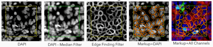
    Today, let’s explore three different approaches to segmenting cells in samples stained with various multiplexed fluorescent assays.

    Starting with the challenge: Precision in Complexity

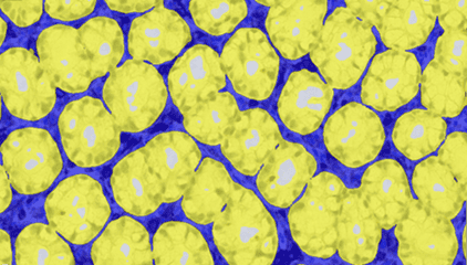
    In tissue pathology, multiplex tissue images have become crucial, allowing us to observe each cell’s location and understand its complex features. Yet accurately and automatically defining each cell’s outer boundary for analysis proves challenging. The reason for that is that tissues contain a heterogeneous mix of cells, each of their own shape and size. Commonly used methods for cell segmentation only focus on nuclei, but those often fall short in precisely defining cell edges.

    Let’s compare the methods: Traditional vs. A.I.-Powered 

    In this study, Daniel Winkowski and team used Visiopharm’s image analysis software to explore three different approaches to segment cells in samples stained with different multiplexed fluorescent assays:

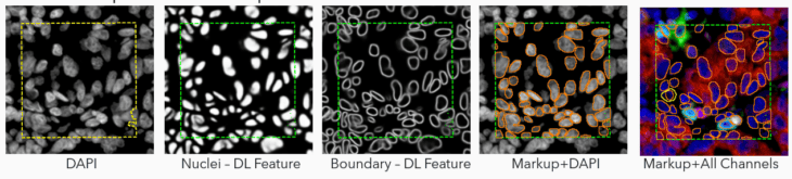
    1. Traditional Image Analysis (Trad): This method uses built-in filtering to enhance objects of interest, relying more on morphometry than intensity. While effective in identifying nuclei based on shape, it’s time-consuming and can struggle with diverse nuclear morphologies.

    2. Ready-To-Use Deep Learning (AI): This approach uses a deep learning algorithm for nuclear signals identification and segmentation. It estimates cell boundaries based on dilation – a no-hassle method but still an estimation. This approach can underperform in identifying cells with clear biomarker staining but lacking a clear nucleus in the plane of section (i.e., CD68 macrophages).

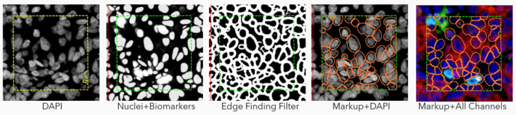
    3. Adapted Deep Learning (AI+): The real star of the show! This method augments the deep learning algorithm to include signals from biomarker channels, assisting in accurately delineating cell boundaries. This blend of nuclei and biomarkers, coupled with an edge-finding filter, offers a more precise approximation of the cell border and assists in finding complex cells (i.e,. macrophages, etc.) where nuclear identification methods fall short.

    Visiopharm offers a complete toolbox for design and customizing your own algorithm but also offers a general nuclear detection algorithm which uses Deep Learning AI to segment nuclei of most morphologies (Ready-to-use AI approach).

    To create the AI+ approach, the team adapted the ready-to-use algorithm to combine its nuclear AI classifier with signals from biomarkers in the multiplex panels to expand the segmentation beyond the nuclei, to the outer cell periphery.

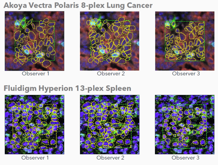
    The performance of these approaches was quantified by comparing them with manual annotations as a ground truth.

    Ground truth annotations were prepared by three human observers on images from two different instrument manufacturers: Vectra Polaris 8-plex lung cancer (Akoya Biosciences) and Hyperion 13-plex spleen (Standard BioTools).

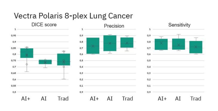
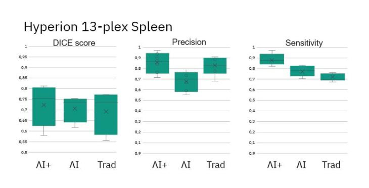
    The AI’s performance was assessed against the ground truth annotations using Precision (a ratio of true and false positives), Sensitivity (a ratio of true positives and false negatives), and DICE Score (a ratio of all true/false, positive/negatives).

    Results: AI+ Takes the Lead 

    After rigorous testing against manual annotations (the ground truth), the results were clear. While all algorithms performed admirably, the AI+ method showed the highest concordance with ground truth annotations. This means a more complete and accurate segmentation of cells across varying multiplex images.

    • DICE Scores for AI+ method show improved performance vs Ground Truth
    • Precision and Sensitivity (measures of object detection) are similar across methods
    • DICE Scores for all methods show similar performance vs Ground Truth
    • Precision and Sensitivity (measures of object detection) are roughly equal across methods

    The Visiopharm has developed a flexible, AI-based strategy enabling comprehensive cell segmentation across various multiplex images.

    • All algorithms performed at a high-level using industry standards including DICE Scores, Precision and Sensitivity. 
    • The Adapted Deep Learning algorithm showed highest concordance with Ground Truth annotations.
    • Performance differences across instruments could be attributed to resolution differences of the instruments, the number of biomarkers in the panel, and differences in images tissue and disease state.
    • Despite annotation differences among human observers, the algorithms perform consistently across the image set.

    Would you like to see how Visiopharm’s software could support your cell segmentation challenges in multiplexed images? Learn more about Phenoplex, a complete workflow for all your multiplexed image analysis needs.

  • Q&A: NovAliX’s journey with Visiopharm’s image analysis software

    Q&A: NovAliX’s journey with Visiopharm’s image analysis software

    Welcome to our latest blog post, where we delve into the evolving landscape of histology and image analysis through an exclusive interview with industry experts from NovAliX – a fully integrated drug discovery CRO based in Strasbourg, France.

    In this insightful conversation, Didier Merciris and Florence Anquetil-Besnard from NovAliX share their experiences in incorporating Visiopharm’s image analysis software into NovAliX’s workflows, and the profound impact it has had on their projects and clients.

    Didier Merciris is a Senior Project Manager at NovAliX where he oversees image analysis for all inflammation related models. Prior to NovAliX, Didier worked for 16 years at Galapagos in the histology department where he was in charge of using image analysis and performing immunohistochemistry and in situ hybridization in different mouse models/therapeutic areas:  
    Inflammation (Osteoarthritis/Arthritis (joints), Osteoporosis (bone), IBD (colon), Lupus (kidney) and fibrosis (liver, kidney, lung).
    Florence Anquetil-Besnard is a Histology Project Manager at NovAliX. After a PharmD and a PhD in Immunology, she began her career in the autoimmune field (rheumatoid arthritis, diabetes) and specialized in histopathology and quantitative image analysis. She is a passionate advocate for digital pathology and loves to solve challenging projects using artificial intelligence (AI) tools. Florence joined the Galapagos team in 2020 to implement the newly formed kidney disease histology area. Her current work within NovAliX also includes oncology projects in diverse organs (brain, pancreas, liver, xenograft tumor…). 

    Visiopharm: Welcome, Didier and Florence. Can you tell us about NovAliX, the services you offer, and how you differentiate yourself from other CROs? 

    Florence Anquetil-Besnard: NovAliX is a fully integrated drug discovery CRO. We support biopharma companies with a range of programs, from initial target identification to the delivery of preclinical candidates. We have seasoned teams in medicinal chemistry and pharmacology, covering a variety of fields – oncology, inflammation, fibrosis, infectious diseases, kidney diseases, and more. We work with a diverse selection of screenings and characterization techniques, including a unique DNA encoded library platform, along with a vast array of biophysical methods, including Cryo-Electron Microscopy (Cryo-EM). Such advanced technologies are rare finds in the market and enhance our research capabilities significantly.

    Didier Merciris: In 2023, our former company, Galapagos, transferred all its discovery resources located in France to NovAliX. This strategic move included integrating a team of highly trained experts, each with over 20 years of experience in fields such as chemistry, protein production, in vivo pharmacology, DMPK (Drug Metabolism and Pharmacokinetics), translational science, and histology. We now offer comprehensive histology services tailored to any requirement within the histology workflow. This ranges from basic tissue processing to advanced staining techniques and in-depth image analyses.

    Visiopharm: What problems were you trying to solve when you started working with us compared with previous analysis software and how did Visiopharm help you overcome those challenges? 

    Didier Merciris: For years, we had been working with an old-fashion image analysis software with limited machine learning capabilities. Developing an image analysis project with this tool required extensive annotations and post-processing steps, which were not only time-consuming, but also yielded unsatisfactory results.

    We were literally stuck due to the software’s limitations, which significantly affected our ability to accurately measure data on slides. This was a major source of frustration in my daily work.

    Florence Anquetil-Besnard: When I joined NovAliX, one of the first things I did was to share my previous experience with Visiopharm. Indeed, I thought the software could significantly improve our capabilities due to its deep learning features; so, we quickly organized a proof of concept with David Mason (Technical Sales Specialist at Visiopharm). The aim was to present him with our most common and challenging issues. We aimed to test the system thoroughly. As expected, Visiopharm, through David’s efforts, was able to provide solutions to all our challenges.

    Didier Merciris: During David’s demonstration, I was really amazed by how Visiopharm easily resolved all our imaging analysis problems. Visiopharm is a complex software to handle, however, thanks to the exhaustive training that we had when we bought the software, it was manageable to learn all its nuances easily.

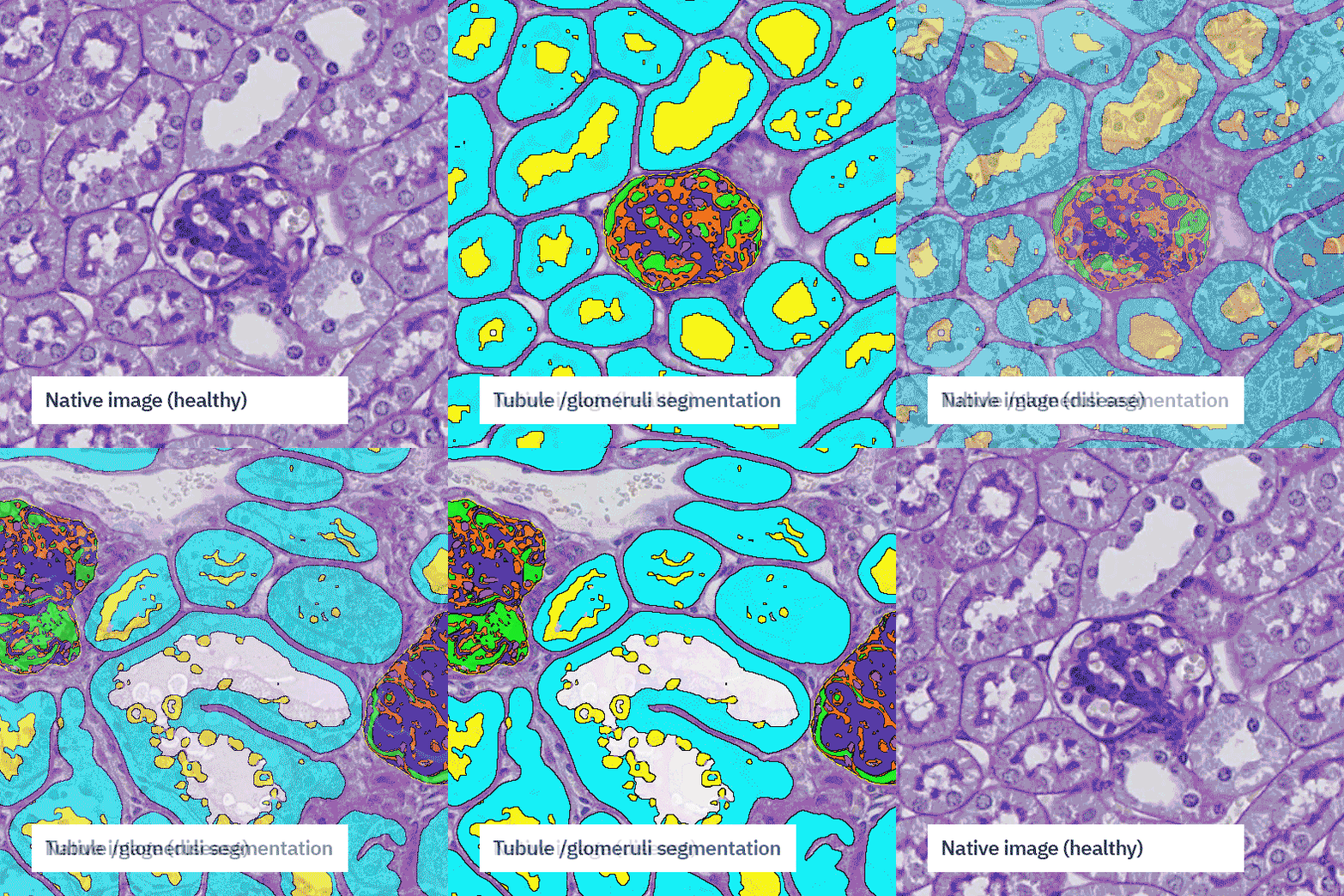
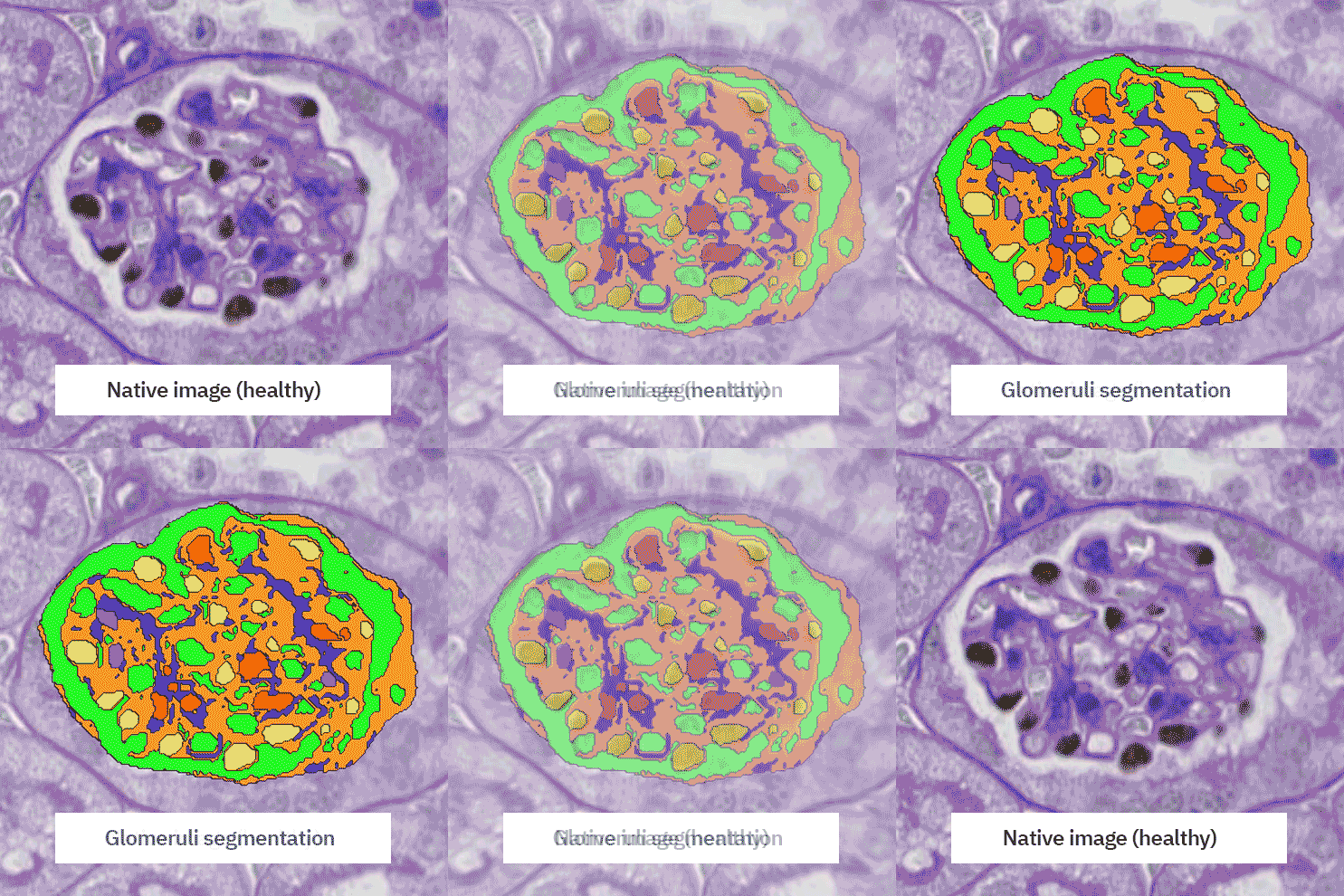
    Florence Anquetil-Besnard: I would say that we are now better equipped to address the complexity of organs. Taking kidney as an example, it is an organ divided into four main compartments: glomeruli, tubules, blood vessels, and interstitium. These parts are independent from each other, but can also be all connected together, posing unique challenges for analysis. Now, with Visiopharm, we can conduct detailed analyses in each area, a task that was significantly more challenging before. For instance, we can independently assess fibrosis in the glomerulus and in the interstitium. This improvement is largely due to the deep learning capabilities of the software.

    Even more impressive, we are not limited to broad compartment analysis; for example, in mice, a single section can contain up to 200 glomeruli. Currently, we can isolate and derive specific data from each glomerulus independently.

    This represents a substantial advancement for our research. Consequently, we can achieve more accurate and representative results.

    Typically, working with a pathologist yields a score for the overall tissue. Now, we can provide a more detailed picture of the tissue section, not just an overall score but insights into specific changes occurring within the tissue.

    Visiopharm: What were you specifically looking for in a solution, how did you go about your search, and what ultimately led you to choose Visiopharm?

    Florence Anquetil-Besnard: For advanced users like me, the support team is also there to bring you even further than what you thought. For example, once I had a big challenge with a complex macrophage staining project involving multiplex techniques. The heterogeneity of the staining was problematic, with intensity varying significantly across samples. In the image analysis community, heterogeneity of fluorescent staining is a nightmare. So, I called the support team, and they guided me through an unconventional use of the software, not something that is taught at first sight. With their help and additional processing steps, we managed to overcome this variability, enabling us to deliver a reliable and robust analysis to our client.

    This experience highlights the importance of not only overcoming technical challenges but also of being able to deliver solid, scientific solutions to our clients.

    Florence Anquetil-Besnard: In the imaging community, it’s crucial to stay updated and leverage the most advanced tools to remain competitive. Our requirement was for a flexible solution capable of meeting the market’s existing standards. While the Oncotopix® Discovery software works for up to 4 markers, the transition to real multiplex analysis requires a more sophisticated approach. The introduction of Phenoplex has significantly simplified the analysis of complex multiplex immunostains.

    Didier Merciris: Another major advantage that influenced our decision was the exceptional support provided by Visiopharm. As a “newbie” to the software, I really appreciate the constant availability of the support team whenever I need them. They respond quickly and efficiently to any queries or issues, which has been incredibly reassuring.

    With Visiopharm, you’re never alone with your challenges; there’s a supportive community, the blog, and a dedicated support team ready to assist.

    Visiopharm: What specific features of our software do you find most helpful?

    Florence Anquetil-Besnard: With Visiopharm, the process is intuitive: start with an image, add annotations, and if opting for deep learning, proceed to train and validate, among other steps. But what is also really nice is the APP Center – it’s like entering a magical world where you can pick an existing app created by someone else, make minor adjustments to fit your needs, and then all of a sudden you have your own customized app, that you didn’t spend time to create, yet still reliable and efficient.

    This flexibility is really helpful to adjust our workflows, offering both time savings and inspiration by exposing us to ideas from other APPs, created by other users in multiple different ways.

    Moreover, the software’s versatility extends beyond just brightfield images, but multiplex fluorescent and special stains, enhancing our research scope. We are empowered to combine different histological techniques, like integrating IHC and ISH, allowing simultaneous investigation of proteins and RNA, or merging special stains like H&E or PAS with targeted IHC staining.

    We can extract as much information as we want from the minimal amount of tissue, which is very helpful when you don’t have a lot of samples to work with, so we can maximize the analysis with just one image.

    Didier Merciris: I used to work with another solution, and to be honest, Visiopharm is faster than the other software. It’s significantly quicker because it can process multiple images concurrently. While the speed can vary based on the user’s IT infrastructure, Visiopharm consistently delivers faster results.

    Compared to our former software, we’ve seen a substantial reduction in processing time, enabling us to deliver data to clients much more rapidly. Another advantage is the user experience, the software was built with an accessible interface that is very easy to use after a little training.

    Visiopharm: Could you share some insights into how the software has added value to your business and your customers?

    Didier Merciris: As you know, the power of Histology lies in its spatial resolution at the cellular level.

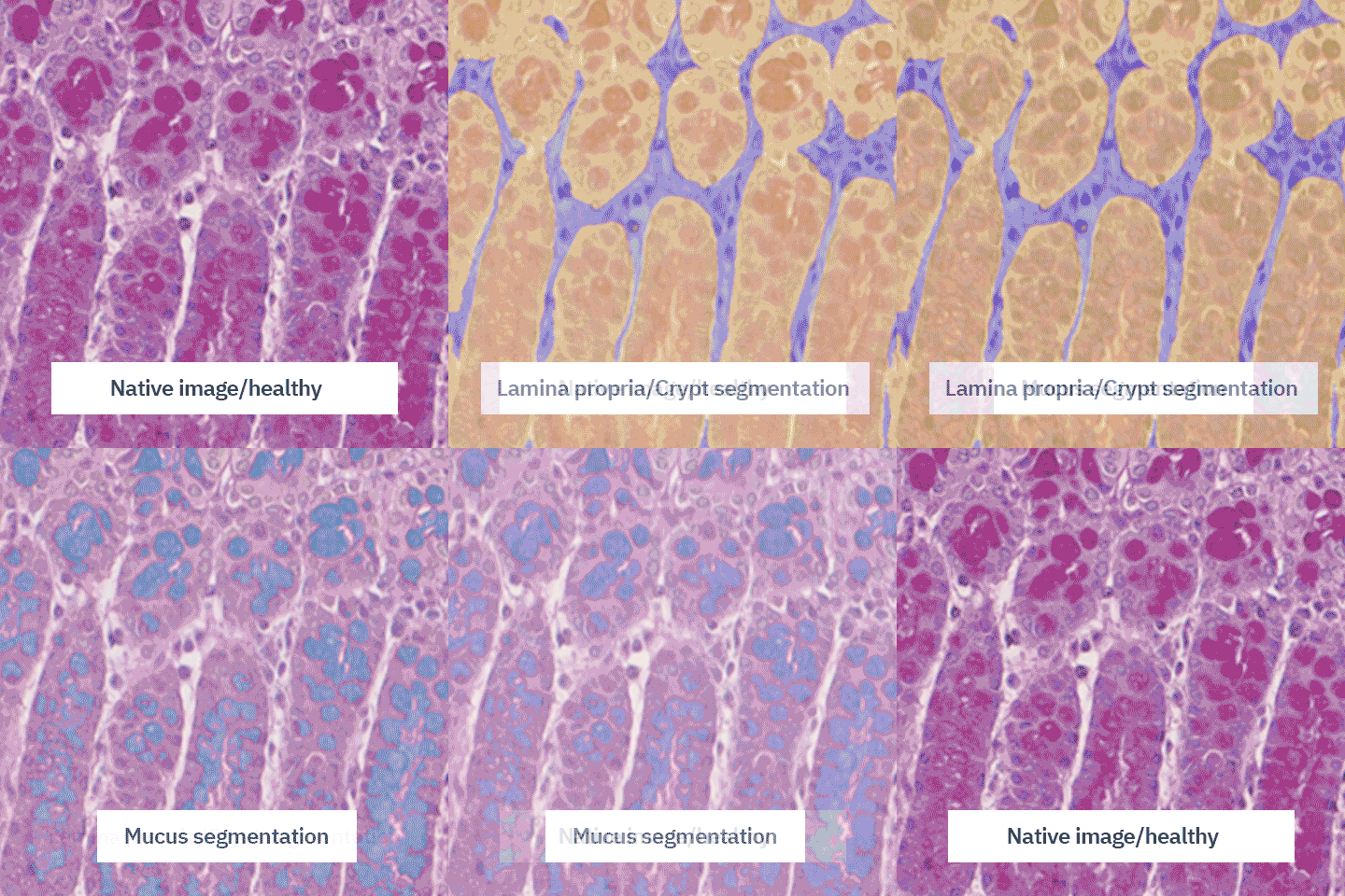
    We had a client interested in quantifying mucus production in epithelial cells from a diseased colon, without considering inflammatory cell infiltration.

    The AI capabilities of Visiopharm significantly aided us in meeting the client’s needs within an extremely short timeframe.

    Moreover, thanks to deep learning, we can tailor our algorithm to work with any staining. Now, if there are any color variations on the slide, we can simply add them to the APP.

    This allows for much greater consistency in our data, which is a significant advantage for us and our clients.

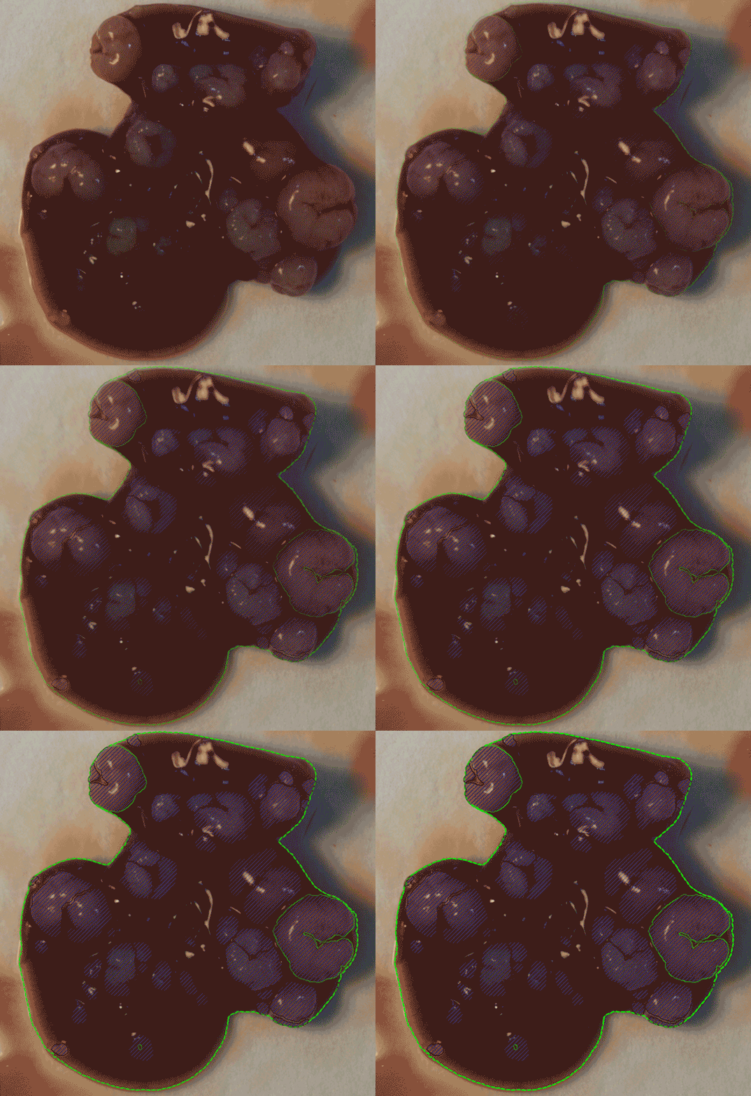
    Liver nodule segmentation

    Florence Anquetil-Besnard: An important aspect is our adaptability as a CRO; it’s not limited to Histology. We can adjust to almost any image. A recent unique project involved working with an image from a camera—not even a microscope, but a camera. We developed a program that automates the counting of tumor nodules in photographs from an ex vivo organ.

    Previously, scientists had to manually count all the nodules. We’ve automated this task. By collaborating closely with our colleagues, we’ve enhanced the reproducibility and reliability of organ assessments. This project was internal, but ultimately, our client benefitted the most because we were able to deliver faster results.

    Stay Ahead – Realize the potential of AI-driven precision pathology.

    In conclusion, NovAliX’s integration of Visiopharm’s image analysis software has significantly transformed their histology and image analysis workflows, offering substantial benefits to their services and clients:

    • Enhanced research capabilities: The adoption of deep learning and artificial intelligence within Visiopharm has empowered NovAliX to overcome previous limitations in image analysis, enabling more detailed and accurate data extraction from tissue samples.
    • Customization and flexibility: The ability to customize algorithms and adapt to a variety of image types has allowed NovAliX to tailor their services to the specific needs of their clients, enhancing the adaptability and reach of their offerings.
    • Overall business value: By leveraging Visiopharm’s technology, NovAliX has not only enhanced their service quality but also solidified their position as a cutting-edge CRO, capable of delivering high-quality, reliable results to their clients in the drug discovery industry.

    Curious to hear more about Visiopharm software? Contact us here.

    Reach out to NovAliX to hear more about their services here.

  • Phenoplex v3: Revolutionizing multiplex image analysis with advanced spatial neighborhood capabilities  

    Phenoplex v3: Revolutionizing multiplex image analysis with advanced spatial neighborhood capabilities  

    We are excited to launch Phenoplex v3, the latest iteration of our complete workflow for all your multiplex image analysis needs.

    With an emphasis on empowering researchers with unprecedented insights into spatial relationships, Phenoplex v3 introduces groundbreaking features for spatial neighborhood analysis, allowing users to define custom radii and quantify and visualize specific cellular neighbors. 

    Come visit us at SITC booth #716 to see a live preview of the new spatial neighborhood analysis features. 

    Key new features of Phenoplex v3 putting spatial analyses at your fingertips: 

    Proximity Profiling:  

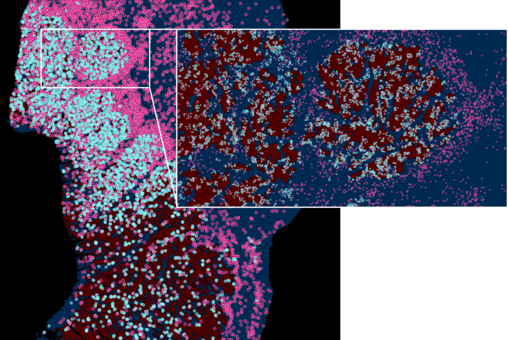
    Phenoplex v3 puts control in the hands of researchers with the ability to define cell population targets and Regions of Interest for analysis. Cell neighbors are selected and custom distance radii around targets can be set. Users can now select relevant cell populations for analysis and display targets and neighbors on their images interactively. This novel feature empowers users to tailor their analyses with unparalleled precision, ensuring that the spatial relationships within user-defined areas are thoroughly examined – spatial biology hypothesis review at a new level.

    Review cellular neighbors on your whole slide images with ease
    Analyse neighboring cells in individual Regions of Interest (ROIs)

    Cross-Population Distance Analysis:  

    Unlock the potential of your multiplex imaging data with Phenoplex v3’s spatial distance analysis. Explore intricate cellular interactions and uncover hidden patterns within tissues and across samples. The intuitive interface simplifies the process, making distance analyses accessible both to experienced users and newcomers, for distances of cell populations to each other cells or to specific Region borders.

    Seamless Workflow Integration: Efficiency Redefined  

    Phenoplex v3 seamlessly integrates into your existing workflow, streamlining the analysis process and saving valuable time. The intuitive user interface, coupled with user-friendly features, ensures a hassle-free experience from data input to insightful results.  

    Paving the Way for Future Discoveries: Stay Ahead in Research  

    With Phenoplex v3, researchers can stay ahead in the dynamic field of multiplex image analysis. The innovative features offered by Phenoplex v3 open new avenues for exploration, helping scientists make groundbreaking discoveries and contribute to the advancement of knowledge.  

    Visiopharm remains committed to providing cutting-edge solutions that empower researchers to unravel the complexities of biological systems. Phenoplex offers you:  

    • A guided bi-directional workflow tailored for setting phenotypes with continuous QC and review of results.  
    • Advanced fully interactive Data Exploration, using t-SNE, Scatter and Box-plotting capabilities along with a cell gallery and cell locations across multiple images  
    • Powerful pre-trained nuclear detection APPs for multiplex immunofluorescence and IMC (imaging mass cytometry).  
    • Easy-to-use channel management tools to QC images and review biomarker localization. Group your channels of interest in multiple meaningful color channel groups to quickly toggle between panels.  
    • A workflow to review and set image object thresholds for each biomarker manually or automatically.  
    • A novel graphical co-occurrence matrix overview to review biomarker double positivity combinations; users with domain knowledge now have an intuitive way to spot unexpected combinations.  
    • Powered by Visiopharm’s best-in-class image analysis platform, combining 20 years of tissue expertise with innovative state-of-the-art technology.  

    Janusz Franco-Barraza, MD, PhD, Research Assistant Professor (E. Cukierman Lab) & Manager of Spatial Immuno-Proteomics Facility at Fox Chase Cancer Center:

    Visiopharm’s Phenoplex workflow allowed me, as a cancer biologist, to effectively scrutinize high-plex immunofluorescence images of pancreatic cancer, investigating a large panel of markers and uncovering the spatial relationships of cell populations, despite being a naive user. This user-friendly platform bridges the gap between basic and translational research, representing the future of cancer cell biology.

    Dr. Fabian Schneider, Visiopharm Research Product manager:

    We’ve learned from our customers and partners that image analysis of highplex assay (>10 channels) has advanced needs that range from how to work with channel management tools, to image object thresholding and clear presentation of biomarker combinations as phenotypes. We have partnered with our users to develop an end-to-end product solution for the analysis of highplex assay imagery. Phenoplex provides researchers with a workflow from dataset management, image object generation using powerful AI APPs, development of channel rules for positivity thresholds, bi-directional data interaction using advanced plotting capabilities, and now advanced spatial neighborhood analysis. With this, we enable researchers, irrespective of their experience with image analysis software or their programming skills, to generate a phenotyping data library and to deep dive into the biology of their datasets.

    For more information about Phenoplex, please visit our website or book a demo of the new features.