Despite the ever-expanding array of technological advancements, image-based research remains limited by the lack of integration between analysis applications and the image management systems central to organizing study data. The disconnect creates bottlenecks in running analysis applications at scale and limits the impact that powerful image analysis results can have as a component of high-throughput research.
Streamline digital precision pathology
That is why Visiopharm, the leader in precision pathology software, and Proscia, a leader in digital pathology image and workflow management, have joined forces to integrate image analysis and image management in a single unified solution. The solution enables life sciences organizations to streamline workflows and accelerate breakthroughs by better leveraging computational image data in research and discovery.
Integrating two powerful platforms
Announced in March, the integrated solution connects Visiopharm’s AI-image analysis suite with Proscia’s Concentriq® for Research image management solution. The integration allows scientists and pathologists to automatically stream images from Concentriq into the VIS suite for analysis and then push results back into Concentriq, where they can be visualized and incorporated into ongoing studies by collaboration partners around the globe.
With a library of over 100 image analysis applications ready to apply to tissue data plus the possibility to modify and create new applications, the Visiopharm software suite enables virtually endless possibilities when it comes to discovering cells, tissues, regions, or any other important piece of data within whole slide images.
Concentriq for Research is an open digital pathology image management system used by leading life sciences organizations to generate big data insights from tissue-based research. It serves as a central hub for managing single or multi-site studies, connecting image analysis applications, information systems, and scanners to streamline image-based workflows. Through the integration, Visiopharm and Proscia are creating a connected digital ecosystem that eliminates data silos and facilitates the introduction of analysis results into routine research. As a result, users can make faster, more informed decisions by harnessing the power of integrated image analysis at scale.
The advancement of digital pathology has led to revolutionary improvements in patient care and medical research. The cellular microenvironment is complex, but the ability of researchers to interrogate it has improved with the development of technologies such as Imaging Mass CytometryTM (IMCTM, Fluidigm) and high-plex fluorescent staining panels (e.g. Akoya or Ultivue). However, increases in the amount and complexity of the data that these technologies yield has brought on a new challenge: developing methods capable of detecting meaningful differences in the datasets.
Introduction to multispectral imaging and analysis
Dr. Heather Stevenson is Associate Professor at the University of Texas Medical Branch and Director of Transplantation Pathology. Her primary research focus is hepatic immunology and how dysregulation of the immune response leads to fibrosis development. She is researching how hepatic macrophage phenotypes determine immune activation and reaction to liver injury. In her latest study, she investigated the phenotypes of intrahepatic macrophages in patients with different types of chronic liver disease.
To obtain a full picture of the microenvironment and to distinguish multiple cell populations, multiple specific biomarkers are needed. To safely assess those biomarkers, various new techniques have been developed, like flow or mass cytometry. Typically those methods dissolve the local context, so that localizations and relations of the cell populations cannot be measured and valuable information is lost. Flow cytometry is unable to visualize multiple antigens in the context of hepatic architecture, and also requires fresh human tissue. It cannot be used for FFPE tissue, which is often more readily available. Single-cell RNA sequencing and mass cytometry can detect multiple markers on intrahepatic macrophages, which is an improvement compared to flow cytometry, but neither can preserve the hepatic architecture or detect locations of the identified cell populations.
Additionally, macrophages are difficult to work with in cell culture, as they can change phenotype when cultured and are not easily isolated from human liver tissue. Mouse models cannot be used for phenotyping intrahepatic macrophages, as they do not adequately replicate the long-term chronic diseases that commonly affect humans, including most liver diseases.
The analyses that most closely replicate the in vivo hepatic microenvironment are in situ methods, rendering multispectral imaging of the tissue slide the method of choice for this type of investigation. Multiplex image analysis is commonly used in immunology research, very prominently in immune-oncology, and also in the investigation of immune cell types in the liver. It also allows to maximize the information collected from a single tissue slide. This is important, since the liver biopsies are being collected in an invasive procedure and are limited, so more information may allow to personalize treatment in the future depending on cellular phenotypes present.
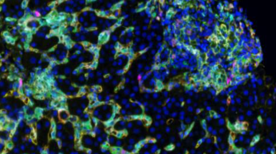
In a recent project, Dr. Stevenson used spectral imaging microscopy (Vectra 3, Akoya Biosystems) to phenotype intrahepatic macrophages in patients with different chronic diseases of the liver, including chronic hepatitis C (HCV), nonalcoholic steatohepatitis (NASH) and autoimmune hepatitis (AIH). Dr. Stevenson and her team analyzed their image data using Visiopharm’s multiplex phenotyping module, which provides efficient and reproducible analyses of high-dimensional multiplexed images.
The technical challenges of working with liver tissue
The first challenge of the study was the high background autofluorescence that liver tissue tends to show due to the high amount of fluorophores. This usually requires specialized equipment to perform spectral unmixing (done by the Vectra System), which removes background autofluorescence from the meaningful data. Following application of the multiplex staining protocols, a large volume of data is generated, especially if researchers are using batch analysis with multiple patients per group. The Visiopharm software robustly copes with the IF images obtained from the Vectra System and facilitates these big data analyses increasing efficiency and reproducibility.
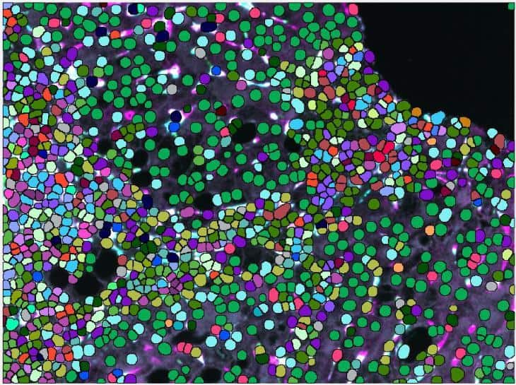
A second challenge presented by this liver tissue study was the difficulty of macrophage segmentation, which can be problematic due to the morphometry of macrophages. It is especially demanding in liver tissue since macrophages are not the primary cell type and their boundaries are often obscured when they are among hepatocytes and epithelial cells.
Very important to the phenotype assessment was also to use an unsupervised approach to deliver the spectrum of phenotypes for all investigated chronic liver diseases and be able to compare those. Using less advanced image mining tool, this step can easily introduce bias to the data and tends to be rather tedious, keeping in mind, that there were 32 possible combinations of macrophage subpopulations to determine per patient and compare between the groups.
Visiopharm’s strategies to overcome liver tissue challenges
Visiopharm’s pretrained AI analysis tools can accurately segment macrophages without additional training of the algorithm, which is an advantage compared to other deep learning platforms that need to be trained from scratch on large annotated datasets. This can take time and also create bias, depending on the training dataset that is used and especially if unsufficient training data is available. The VIS AI tools were able to easily identify the cells for analysis without the need for further training.
The phenotype module of Visiopharm’s software provides the exact framework for analyzing any dataset, by offering the possibility to take an agnostic approach and allowing the data itself to dictate the downstream results, rather than having them be influenced by a priori expectations. “We were quite surprised at the accuracy and precision of the automated phenotyping and the simplicity in using the software,” Dr. Stevenson says of Visiopharm’s phenotyping module. “One of the really interesting findings that is consistent with our overall thoughts is that CD68 and CD163 are not found on the same cells, despite being fairly interchangeable in most of the macrophage literature.” This suggests, that the Visiopharm software had detected differences in cell phenotypes that were not previously known to researchers, which is revolutionary not only for researchers studying liver diseases, but also for all scientists working in the field of immunology.
Visiopharm’s software also provides advanced visualizations to investigate, compare and confirm the results of the analysis. Plots of the dimensionality-reduced data by the t-distributed stochastic neighbor embedding (tSNE) algorithm showed clear differences between the groups of Dr. Stevenson’s study. The data points from each individual case were mapped to the same coordinates, making comparisons between the patients with different liver diseases simple and efficient.
Phenotype color coding was matched between the tSNE plots and the image annotations, which allowed the images to be easily referenced. The phenotypic profile plots showed relative expression levels of different macrophage phenotypes across patient groups in a single simple graphical representation. These software features greatly facilitate what is typically an extremely complex and time-consuming analysis.
Future directions following the initial study
The results of this study allowed researchers in Dr. Stevenson’s lab to expand their panel to other immune cell types, as well as more specific immune cell subtypes. “We can now probe the liver for specific macrophage subsets in addition to other cell types to better understand mechanisms of liver disease and disease signatures,” explains Dr. Stevenson.
Spectral imaging microscopy followed by Visiopharm’s software image analysis is a novel method of phenotyping intrahepatic macrophages in patients with different liver pathologies and has the potential to change the current understanding of intrahepatic macrophages, as well as the way that patient liver biopsies are evaluated in the future.
More information is available by attending this free webinar.
Visiopharm is very happy to announce that Ralf Huss, M.D, Ph.D. has been appointed as the chairperson of their scientific advisory board. As the new academic leader of the advisory board, he brings a wealth of scientific and industry leadership experience in digital pathology.
Currently, Ralf Huss is a Professor of Pathology and the managing deputy director of Pathology and Molecular Diagnostics at the University Hospital in Augsburg (Germany), where he also leads the Center for Digital Medicine. He is board-certified in anatomical, experimental, and molecular pathology with more than 30 years of experience in histopathology, immunology, cancer research and oncology.
“The interplay between the scientific community and companies like Visiopharm represents an essential partnership that helps drive advances in digital pathology. In particular, the need for communication between these two worlds is evident during these extraordinary times. For my part, I am looking forward to developing an international board of advisors that ensures Visiopharm continues to lead the field with technologies and partnerships that bring value to the field.”
His advisory role will begin on May 1, 2020, and among his responsibilities will be structuring the advisory board with a particular focus on guidance in the areas of clinical pathology and quality management of pathological workflows.
“I couldn’t be more pleased to have Ralf join as chairperson of our scientific advisory board. Building a strong advisory board ensures that we maintain close ties to the community and create solutions that pathologists need. With his practical experience and connections in the digital pathology field, Ralf will be instrumental in guiding the future success of Visiopharm.”
Visiopharm is pleased to announce that Dirk Vossen Ph.D. has joined the company as its new Chief Diagnostics Officer.
Dirk brings extensive experience as a global leader in digital pathology, specialized in cancer tissue diagnostics and clinical workflows. His track record of creating value through innovation in digital and computational pathology spans the entire range of development, from ideation through validation and certification of medical devices, as well as commercialization strategies.
Based in The Netherlands, Dirk will be leading Visiopharm’s clinical diagnostics programs, with cross-functional responsibilities that support the company’s continued expansion into diagnostic and clinical applications of digital pathology.
“I have worked with Dirk as a partner, and have admired not only his deep understanding of the digital pathology market, but his innovative leadership and drive to create solutions that propel the industry forward and ultimately improve the care that cancer patients receive,” says Michael Grunkin Ph.D., CEO, and co-founder of Visiopharm. “That’s why I am delighted to welcome him to Visiopharm and look forward to his contributions as we continue on our journey to create precision pathology solutions that elevate the industry standards for diagnostic and clinical image analysis.”
As the co-founder of Philips Digital and Computational Pathology business, Dirk brings over a decade of experience in building digital pathology solutions, including the development of whole slide image scanners, image management systems, computational pathology applications as well as driving the strategies and execution of clinical programs. A key addition to the management team, Dirk will orchestrate the successful development and commercialization of clinical products that put the latest advances in tissue mining into the hands of pathologists.
“It’s an extraordinary time to join Visiopharm, with digital pathology becoming mainstream and breakthrough AI applications that will elevate the quality of data that pathologists use to make critical decisions for patients,” he said. “I am eager to be a part of this next phase of the company’s growth.”
Visiopharm and Proscia® Partner to Deliver AI-Enabled Solutions that Advance Precision Medicine
Collaboration unifies image analysis and image management in a single solution for realizing the full promise of computational pathology data
Visiopharm, the leader in precision pathology software, and Proscia, a leader in digital pathology image and workflow management, have entered into a multi-year, strategic partnership to deliver deeply integrated AI-enabled solutions that drive breakthroughs in translational research and advance precision medicine. In addition to joint product integration, Visiopharm and Proscia have established a global reseller and support agreement to accelerate the delivery of these combined offerings to the market.
The shift towards precision medicine has led to the rapid adoption of digital pathology across translational research, as new computational applications have the potential to unlock hidden insights that drive discoveries into breakthrough therapies. Despite this digitization, research organizations are struggling to incorporate computational data into the modern digital workflow due to the siloed deployment of isolated applications across image analysis and image management.
Visiopharm and Proscia have joined forces to deliver a best-in-class unified solution that enables research organizations to better leverage computational image data. Available today, the integrated solution connects Visiopharm’s VIS AI-image analysis suite with Proscia’s Concentriq® image management platform, enabling high-throughput research organizations to realize the full potential of their digital pathology data. This integration allows scientists and pathologists to automatically stream images from Concentriq into the VIS suite for analysis and push results back into Concentriq, where they can be visualized and incorporated into ongoing studies.
“We are impressed with the innovation, drive, and depth of understanding Proscia brings to the field of image, data, and workflow management,” said Visiopharm CEO Michael Grunkin. “This collaboration will allow each of our companies to focus on our respective core competences. Essentially, that means we can use our resources to keep innovating and pushing the ‘boundaries of possible’ in our respective fields and continue to deliver relevant and future-proof solutions that span the entire scope of precision pathology collaborative workflows.”
“Our strategic partnership with Visiopharm brings together industry-leading technology capabilities and a strong track record of customer success,” said David West, CEO of Proscia. “Through our joint solution, we’re helping our customers adopt an increasingly complex and data-driven approach to pathology that will drive research breakthroughs and lead to improved patient outcomes.”
Enabling better cancer diagnostics via artificial intelligence-driven solutions
Agilent Technologies Inc. (NYSE: A) and Visiopharm today announced that they have entered a long-term collaboration agreement. The announcement is happening at the USCAP 109th Annual Meeting being held on February 29 through March 5, 2020, in Los Angeles, CA.
Based on a joint vision of end-to-end quality and standardization of tissue diagnostics, the partnership will address the currently unmet needs of pathology labs around the world. The companies’ shared goal is to provide specific technologies, products, and services that will improve the standardization of pathology labs and accelerate accurate diagnoses.
Agilent and Visiopharm will co-market Visiopharm’s portfolio of artificial intelligence-driven pathology solutions.
“This partnership is an exciting step in our shared commitment in the fight against cancer,” said Simon Østergaard, Agilent vice president and general manager of the company’s pathology group. “Agilent’s market-leading portfolio of pathology staining management solutions combined with Visiopharm’s digital interpretation solutions will help facilitate improved patient care and diagnostic accuracy. Together we will tackle some of the most critical challenges faced by pathology labs and pave the way toward the fully digitalized lab of the future.”
“The transformation into next-generation precision pathology is important to realize the full potential of tissue pathology in this dawning era of personalized medicine,” said Michael Grunkin, CEO of Visiopharm. “This transformation requires a holistic approach to standardization along the entire diagnostic journey from biopsy to diagnosis. We are excited to be working with Agilent on precisely that. With their complementary product portfolio and longstanding legacy of innovation and quality in this field, we see a very strong match.”
The co-marketing relationship will result in offerings that will improve the standardization of pathology labs and accelerate accurate diagnoses for their patients. Digital pathology is the future, although many labs have waited to adopt it due to the lack of choices currently available and the expense of adoption. This effort will enable labs to gradually adopt digital technologies in a flexible and scalable manner.