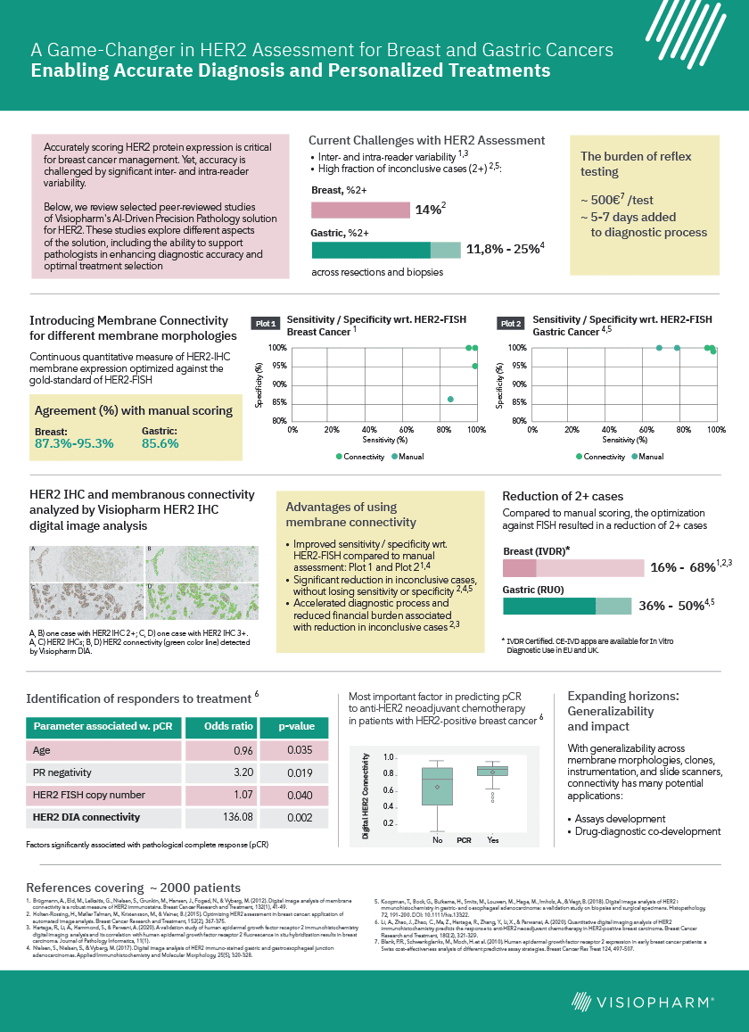
Explore our decade-long journey and research collaboration with international experts. In this infographic, we highlight the potentially transformative impact of our AI-driven Precision Pathology solution on HER2-targeted treatment strategies for breast- and gastric cancer patients. This IVDR-cleared methodology is currently used in clinical practice for decision support when reading HER2-IHC assays. It enhances diagnostic accuracy, is optimized for concordance with HER2-FISH, significantly reduces the need for reflex testing, and has been shown to identify responders to anti-HER2 neoadjuvant chemotherapy.
We will soon release a whitepaper on this subject, which you can sign up to receive by submitting your email further down this page.
Whitepaper sign up
Share your email to get notified when the whitepaper is published.
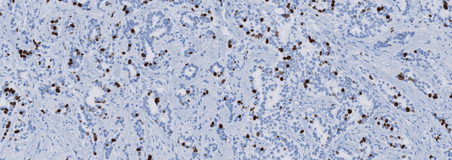
Immunohistochemical staining of tissue slides is a powerful tool that can provide pathologists and clinicians great insight for diagnosis, prognosis, and treatment of many disease states, particularly cancer.
However, like most tools that offer such significant benefits, it doesn’t come without challenges. The quality of staining in immunohistochemistry is an essential factor that may limit its effectiveness as a clinical tool. To correct the quality issues, it is vital to first understand how quality issues arise.
Stain quality can be described in terms of a signal-to-noise ratio (SNR), where signal refers to the level of expected epitope staining, and noise is the background component that results from cross-reactions with undesired epitopes. Insufficient SNRs are commonly the result of using poor or nonrobust antibodies and improper calibrations, the result of which mostly are false negative tests. Careful consideration for proper controls and adherence to quality control within the lab performing the staining protocol must be a priority to achieve optimal stain quality.1
An underlying reason for the variable quality being delivered by labs is the inherent complexity of an immunostaining protocol. Not only are protocols constantly being optimized, but with over 50 steps, each with multiple options, the staining protocol can have millions of variations. This has made standardization difficult.
One study conducted by the Nordic Immunohistochemical Quality Control (NordiQC) found an error rate in staining of almost 30% in labs participating in their study.
One study conducted by the Nordic Immunohistochemical Quality Control (NordiQC) found an error rate in staining of almost 30% in labs participating in their study.2 Dr. Vyberg, an expert in immunohistochemical quality control, noted that one reason for this high error rate is the use of in-house or “home-brew” kits rather than ready-to-use kits, which has direct consequences for treatments. Labs avoiding ready-to-use kits to save money on the front end could actually end up costing hospitals more in the long run.
An example is in the detection of the HER2 protein in breast cancer, one study calculated that every $1 labs saved by using in-house kits for HER2 rather than ready-to-use kits cost the hospital $6.3 However, quality assessment organizations have no authority to enforce underperforming labs to change their protocols.
This is not to suggest that all ready-to-use products are error-proof; internal standardization within each operating lab is critical with the use of any kit. Additionally, the field must be aware of the constantly evolving methods of staining in order to adopt the best possible protocols.
Early detection for some cancers is critical; delay in diagnosis and treatment increases the death rate considerably.4 Immunohistochemistry has the potential to help in early cancer detection, but first, the issue of stain quality must be addressed. One effort that is currently being made by NordiQC is the transparent sharing of optimal protocols for widespread use on their website. However, awareness and additional efforts are still needed, as solutions to this problem could directly impact cancer patients across the world.
1. Vyberg, M. & Nielsen, S. Proficiency testing in immunohistochemistry—experiences from Nordic Immunohistochemical Quality Control (NordiQC). Virchows Arch. 468, 19–29 (2016).
2. Nielsen, S. External quality assessment for immunohistochemistry: Experiences from NordiQC. Biotech. Histochem. 90, 331–340 (2015).
3. Vyberg M, Nielsen S, Røge R, Sheppard B, Ranger-Moore J, Walk E, Gartemann J, Rohr UP, Teichgräber V. Immunohistochemical expression of HER2 in breast cancer: socioeconomic impact of inaccurate tests. BMC Health Serv Res. 2015 Aug 29;15:352. doi: 10.1186/s12913-015-1018-6.
4. Hanna TP, King WD, Thibodeau S, Jalink M, Paulin GA, Harvey-JonescE, O’Sullivan DE, Booth CM, Sullivan R, Aggarwal A. Mortality due to cancer treatment delay: systematic review and meta-analysis. BMJ. 2020 Nov 4;371:m4087. doi: 10.1136/bmj.m4087.
We’ve given our Customer Center a new look, plus added and consolidated features into one platform. Now you only need to go to one place for support requests, book training, get account info, and download software. The brand-new platform still has the features you know and love, but with new capabilities to help you.
So, what’s new then?
Main page
From the main page, you can access your support cases, APPs, current software licenses, and go to the new Knowledge Base. In the top right, you’ll also find a dropdown menu – but more on that later.
Find and create support cases
By clicking on ‘Show my Cases’ you can see your active cases and follow their progress – it’s also possible to see previous cases in this new feature.
Have a question? Need some additional training? We are happy to help you! Just click ‘Create’ to ask a question, request personalized training, or get technical support and installation.
This is also the place to connect with our Support Team and start a remote session to get online software help.
Here’s how you do it:
See your APPs and licenses
Go to Show my Apps and you will see the list of your purchased APPs. This includes ID, APP version number, and an APP description.
If you’re in doubt about your license – then worry no more. You can now find this information by clicking on Show my Licenses. Here you can find your active licenses and the expiry dates.
Want to do some learning on your own?
We have gathered our training videos and help manual in the new Knowledge Base. Here you can take your image analysis skills to the next level with online training videos, from getting started with the software to diving into advanced APP design.
You’ll find walkthroughs of common examples in our different modules, technical videos on how to do a new installation, and explanations of new software features.
Get the newest edition of the software
Need to update? Go to MyVisiopharm in the top ribbon and unfold the menu – click on Downloads. Here you can download relevant documents or installers. You’ll get to a login page where you need to verify your email through an access code. Click Authorize and type in the Access Pass sent to your e-mail. Voila, you can now download the relevant file(s).
Here’s how you do it:
If you have specific questions, you can always search our new Knowledge Base or access the online manual through the My Visiopharm in the top ribbon.
Despite the ever-expanding array of technological advancements, image-based research remains limited by the lack of integration between analysis applications and the image management systems central to organizing study data. The disconnect creates bottlenecks in running analysis applications at scale and limits the impact that powerful image analysis results can have as a component of high-throughput research.
Streamline digital precision pathology
That is why Visiopharm, the leader in precision pathology software, and Proscia, a leader in digital pathology image and workflow management, have joined forces to integrate image analysis and image management in a single unified solution. The solution enables life sciences organizations to streamline workflows and accelerate breakthroughs by better leveraging computational image data in research and discovery.
Integrating two powerful platforms
Announced in March, the integrated solution connects Visiopharm’s AI-image analysis suite with Proscia’s Concentriq® for Research image management solution. The integration allows scientists and pathologists to automatically stream images from Concentriq into the VIS suite for analysis and then push results back into Concentriq, where they can be visualized and incorporated into ongoing studies by collaboration partners around the globe.
With a library of over 100 image analysis applications ready to apply to tissue data plus the possibility to modify and create new applications, the Visiopharm software suite enables virtually endless possibilities when it comes to discovering cells, tissues, regions, or any other important piece of data within whole slide images.
Concentriq for Research is an open digital pathology image management system used by leading life sciences organizations to generate big data insights from tissue-based research. It serves as a central hub for managing single or multi-site studies, connecting image analysis applications, information systems, and scanners to streamline image-based workflows. Through the integration, Visiopharm and Proscia are creating a connected digital ecosystem that eliminates data silos and facilitates the introduction of analysis results into routine research. As a result, users can make faster, more informed decisions by harnessing the power of integrated image analysis at scale.
The advancement of digital pathology has led to revolutionary improvements in patient care and medical research. The cellular microenvironment is complex, but the ability of researchers to interrogate it has improved with the development of technologies such as Imaging Mass CytometryTM (IMCTM, Fluidigm) and high-plex fluorescent staining panels (e.g. Akoya or Ultivue). However, increases in the amount and complexity of the data that these technologies yield has brought on a new challenge: developing methods capable of detecting meaningful differences in the datasets.
Introduction to multispectral imaging and analysis
Dr. Heather Stevenson is Associate Professor at the University of Texas Medical Branch and Director of Transplantation Pathology. Her primary research focus is hepatic immunology and how dysregulation of the immune response leads to fibrosis development. She is researching how hepatic macrophage phenotypes determine immune activation and reaction to liver injury. In her latest study, she investigated the phenotypes of intrahepatic macrophages in patients with different types of chronic liver disease.
To obtain a full picture of the microenvironment and to distinguish multiple cell populations, multiple specific biomarkers are needed. To safely assess those biomarkers, various new techniques have been developed, like flow or mass cytometry. Typically those methods dissolve the local context, so that localizations and relations of the cell populations cannot be measured and valuable information is lost. Flow cytometry is unable to visualize multiple antigens in the context of hepatic architecture, and also requires fresh human tissue. It cannot be used for FFPE tissue, which is often more readily available. Single-cell RNA sequencing and mass cytometry can detect multiple markers on intrahepatic macrophages, which is an improvement compared to flow cytometry, but neither can preserve the hepatic architecture or detect locations of the identified cell populations.
Additionally, macrophages are difficult to work with in cell culture, as they can change phenotype when cultured and are not easily isolated from human liver tissue. Mouse models cannot be used for phenotyping intrahepatic macrophages, as they do not adequately replicate the long-term chronic diseases that commonly affect humans, including most liver diseases.
The analyses that most closely replicate the in vivo hepatic microenvironment are in situ methods, rendering multispectral imaging of the tissue slide the method of choice for this type of investigation. Multiplex image analysis is commonly used in immunology research, very prominently in immune-oncology, and also in the investigation of immune cell types in the liver. It also allows to maximize the information collected from a single tissue slide. This is important, since the liver biopsies are being collected in an invasive procedure and are limited, so more information may allow to personalize treatment in the future depending on cellular phenotypes present.
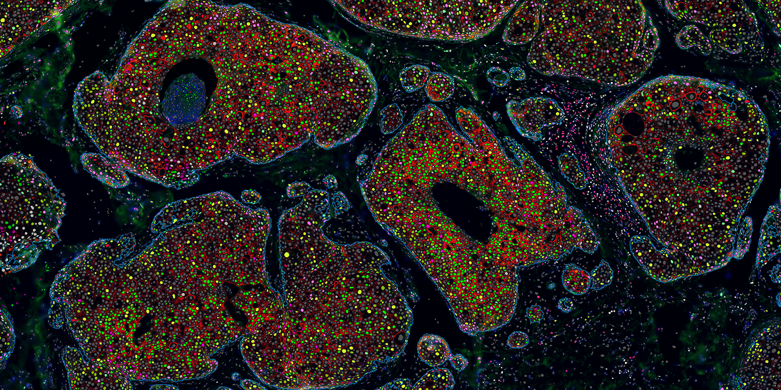
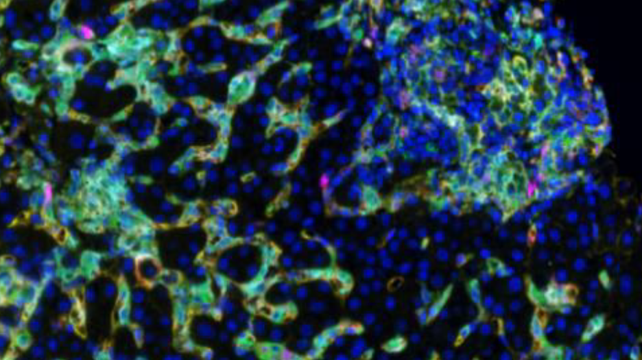
In a recent project, Dr. Stevenson used spectral imaging microscopy (Vectra 3, Akoya Biosystems) to phenotype intrahepatic macrophages in patients with different chronic diseases of the liver, including chronic hepatitis C (HCV), nonalcoholic steatohepatitis (NASH) and autoimmune hepatitis (AIH). Dr. Stevenson and her team analyzed their image data using Visiopharm’s multiplex phenotyping module, which provides efficient and reproducible analyses of high-dimensional multiplexed images.
The technical challenges of working with liver tissue
The first challenge of the study was the high background autofluorescence that liver tissue tends to show due to the high amount of fluorophores. This usually requires specialized equipment to perform spectral unmixing (done by the Vectra System), which removes background autofluorescence from the meaningful data. Following application of the multiplex staining protocols, a large volume of data is generated, especially if researchers are using batch analysis with multiple patients per group. The Visiopharm software robustly copes with the IF images obtained from the Vectra System and facilitates these big data analyses increasing efficiency and reproducibility.
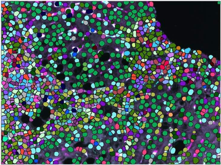
A second challenge presented by this liver tissue study was the difficulty of macrophage segmentation, which can be problematic due to the morphometry of macrophages. It is especially demanding in liver tissue since macrophages are not the primary cell type and their boundaries are often obscured when they are among hepatocytes and epithelial cells.
Very important to the phenotype assessment was also to use an unsupervised approach to deliver the spectrum of phenotypes for all investigated chronic liver diseases and be able to compare those. Using less advanced image mining tool, this step can easily introduce bias to the data and tends to be rather tedious, keeping in mind, that there were 32 possible combinations of macrophage subpopulations to determine per patient and compare between the groups.
Visiopharm’s strategies to overcome liver tissue challenges
Visiopharm’s pretrained AI analysis tools can accurately segment macrophages without additional training of the algorithm, which is an advantage compared to other deep learning platforms that need to be trained from scratch on large annotated datasets. This can take time and also create bias, depending on the training dataset that is used and especially if unsufficient training data is available. The VIS AI tools were able to easily identify the cells for analysis without the need for further training.
The phenotype module of Visiopharm’s software provides the exact framework for analyzing any dataset, by offering the possibility to take an agnostic approach and allowing the data itself to dictate the downstream results, rather than having them be influenced by a priori expectations. “We were quite surprised at the accuracy and precision of the automated phenotyping and the simplicity in using the software,” Dr. Stevenson says of Visiopharm’s phenotyping module. “One of the really interesting findings that is consistent with our overall thoughts is that CD68 and CD163 are not found on the same cells, despite being fairly interchangeable in most of the macrophage literature.” This suggests, that the Visiopharm software had detected differences in cell phenotypes that were not previously known to researchers, which is revolutionary not only for researchers studying liver diseases, but also for all scientists working in the field of immunology.
Visiopharm’s software also provides advanced visualizations to investigate, compare and confirm the results of the analysis. Plots of the dimensionality-reduced data by the t-distributed stochastic neighbor embedding (tSNE) algorithm showed clear differences between the groups of Dr. Stevenson’s study. The data points from each individual case were mapped to the same coordinates, making comparisons between the patients with different liver diseases simple and efficient.
Phenotype color coding was matched between the tSNE plots and the image annotations, which allowed the images to be easily referenced. The phenotypic profile plots showed relative expression levels of different macrophage phenotypes across patient groups in a single simple graphical representation. These software features greatly facilitate what is typically an extremely complex and time-consuming analysis.
Future directions following the initial study
The results of this study allowed researchers in Dr. Stevenson’s lab to expand their panel to other immune cell types, as well as more specific immune cell subtypes. “We can now probe the liver for specific macrophage subsets in addition to other cell types to better understand mechanisms of liver disease and disease signatures,” explains Dr. Stevenson.
Spectral imaging microscopy followed by Visiopharm’s software image analysis is a novel method of phenotyping intrahepatic macrophages in patients with different liver pathologies and has the potential to change the current understanding of intrahepatic macrophages, as well as the way that patient liver biopsies are evaluated in the future.
More information is available by attending this free webinar.
CIO Applications recently sat down with our founder and CEO Michael Grunkin. The technology print magazine focuses on usage of various tech start ups. In the latest issue the spotlight was placed on the marketplace of machine learning providers and the difficulties in zeroing in on the best. To address this, CIO Applications took up the challenge and located the “Top 25 Machine Learning Solution Providers – 2019”.
Here is the full interview from CIO Applications where CEO Michael Grunkin, shared his insights into how AI and machine learning-enabled image analysis software has demonstrated the potential to make a real difference in tissue-based research and diagnostics.
Augmenting Pathologists—Image Analysis with AI
Clinicians rely heavily on tissue samples that are interpreted by pathologists under a microscope. This practice has been in place for hundreds of years and is now ready to be automated, as leading quantitative digital pathology solution provider, Visiopharm, applies AI and Machine Learning in their image analysis software. Digitized pathology images, known as Whole Slide Images (WSI), represent several challenges to obtain meaningful, reproducible, and standardized analysis.
Interesting advances in technology usually always happens at the intersection between different fields of science and technology. Diagnostic quantitative digital pathology is no exception: it lies firmly at the intersection between computer science, image analysis, and applied mathematics. I was a technical founder of two earlier diagnostic companies based on image analysis. This was a valuable learning experience that showed the enormous potential diagnostic image analysis has toward providing both data quality and automation. It later became clear that there is a huge unmet need in the field of pathology.
Pathology relies heavily on manual reading and interpretation of biological tissue structures. With subjective and error-prone manual processes, diagnostic digital pathology has an enormous potential to automate and improve tissue-based diagnostics. This is becoming increasingly important in an era of new targeted cancer therapies. These treatments have the potential to significantly improve the prognosis for cancer patients. The driving vision behind Visiopharm, which my co-founder Johan Dore and I founded back in 2001, is to support pathologists and scientists with the tools to identify the right treatment, for the right patient, at the right time.
What are some trends that you see emerging in pathology, and how is Visiopharm leveraging those?
The advent of WSI has enabled the market for quantitative digital pathology both in diagnostics and research. WSI allows you to digitize an entire microscope slide at very high resolution and view it from a monitor. Digital Pathology helps scientists and pathologists to replace the traditional microscope, and assess important cellular and morphometric structures more efficiently directly on screen.
The ability to digitize slides is just the first step in the journey toward automated precision pathology. The manual assessment of complex biological tissue structures and biomarker response often represents a formidable cognitive challenge, especially with a whole new generation of biomarkers that are used to characterize the tumor micro-environment. Over the years, we have been able to understand, identify, and quantify the error sources from biopsy to data. We have taken a radical approach to simplify and improve the entire process for pathologists. We use machine learning and AI technology to mitigate or eliminate these error sources and achieve higher interpretive accuracy of tissue data.
Visiopharm’s main focus is cancer research and diagnostics. As we speak, there are more than 600 new immunotherapies in late stage development.This new generation of cancer treatments presents new challenges for pathology. Immunotherapies are expensive targeted therapies, meaning that only a sub- population of patients will respond to the treatment.This is where Visiopharm makes a difference. Our image analysis workflows and algorithms improve the interpretive accuracy of some of the most challenging and time-consuming manual pathology reads.Our technology can provide decision support for pathologists and scientists that are tasked with making important diagnostic, research or business decisions based on tissue data.
How has Visiopharm’s Oncotopix® and AI been able to imprint its exclusivity in the pathology market?
The simplicity in workflow, combined with the flexibility and power of the software, enables our customers to easily configure new diagnostic or research APPs without the need for programming skills. In particular, our commitment to data quality, has made us one of the most widely-adopted solutions in the market.
Without consistent good stain quality, it is often not possible to make an accurate diagnosis. Visiopharm has been able to use our image analysis platform to find new and effective ways to measure the variability in stain quality. We are working closely with EQA organizations to provide Qualitopix™, our platform that will effect change and significantly impact the standardization of tissue diagnostics.
Lastly, we have invested significantly in integrating to existing digital infrastructure, including Image Management, Lab Information, and PACS systems. Our solutions now plug directly into existing workflows and augment the pathologist with APPs for precision pathology.Could you cite a case study describing how you enable clients to overcome hurdles and obtain desired outcomes with your innovative digital pathology solutions?
We recently supported Karolinska Institute in Stockholm on a research project to understand the magnitude of manual reading errors throughout the diagnostic process. The researchers used Ki-67, a biomarker for distinguishing between two sub-types of breast cancers, where the treatment selection depends on the classification of the patient into one of these two sub-types. The study demonstrated a 30 percent manual misclassification rate between these cancer types. Visiopharm’s software significantly reduced this error.
We also recently finished installation of our Oncotopix® Dx for breast cancer in hospitals throughout Denmark.
Some of the most notable results, was considerable time and money savings on reflex molecular testing for inconclusive breast cancer cases. For a large fraction of patients, we could completely eliminate the need for the molecular test. This saves money for laboratories, but more importantly, it greatly reduced the waiting time for these patients.
What does the future hold for your organization?
Visiopharm continues to demonstrate double-digit year over year growth. This validates both the magnitude of the unmet need in the market, and our ability to address that need effectively. We are selling our solutions to diagnostic pathology labs, biopharmaceutical companies, academic medical centers, and contract research companies that all have similar needs. We see a clear window of opportunity to continue this growth, expand geographically and lead market innovation. With the recent strong financial support from Danish investors, our next phase of growth involves launching our diagnostic technology into the U.S., a market that is already strong for Visiopharm in drug development and research.