Category: Blog

  • DICOM – a Standard Under Construction

    As pathology is going digital our need for standardization is plain as day. For this DICOM (Digital Imaging and Communications in Medicine) is a necessary step on the standardization journey offering independence on scanners and connected software solutions. However, the road to standardization comes with a series of bumps along the way.

    DICOM makes medical imaging information interoperable. Its core function is to integrate image-acquisition devices, PACS, workstations, VNAs and analysis software from different manufacturers such as Visiopharm. People are actively developing and maintaining it to meet the evolving technologies and needs of medical imaging, and it is free to download and use (source: https://www.dicomstandard.org). 


    ​​Not quite there yet


    In my opinion, DICOM isn’t developed enough to be used as an out-of-the-box solution. We’ve taken the first steps in using DICOM in clinical practice and have gotten quite far since I wrote about standardizing digital pathology in 2014. Nonetheless every customer who joins DICOM today will be doing pioneering work and should be aware of it.

    There is more to DICOM than meets the eye, as DICOM doesn’t only consist of the file-format, but also a series of standardized communication components. 

    In recent Connectathons, it has been demonstrated that it is possible to use DICOM to create a vendor-spanning configuration consisting of a scanner, archive, and viewing. However, there is still too little practical experience on how such systems behave and prove themselves in routine operation over a more extended period. 

    ​The missing piece – or at least one of them

    There is still an important component missing to have a complete DICOM scenario, and that is the IHE-PaLM/DICOM compliant communication with the Pathology LIS/LIMS. The standardization committees (WG26 / IHE-PaLM) are just now considering which technology should be the standard in the future. Today a proprietary solution is still needed to enable communication between the scanner and Pathology LIS/LIMS to exchange the metadata.

    From Visiopharm’s point of view, our solution already supports quite a few image formats and DICOM implementations, such as pyramid structure, z-stacks, concatenations, and storage commitment to name a few. However, there are many shortcomings. Alone in reading info from image data, scanners currently only support RGB brightfield scans. Currently, Multispectral/fluorescence images are not supported.

    The potential is there

    All in all, the idea of DICOM is excellent, and the experiences from past Connectathons are quite convincing and promising.  Adding to this, several pioneering hospitals such as VGR in Sweden have implemented a version of DICOM including streaming from VNA.

    However, DICOM in digital pathology still needs time for development, validation, and verification. Every customer who gets involved with DICOM is to be welcomed, as they contribute to advancing the DICOM standard in digital pathology. It is important that they are aware of the given situation and all its consequences if we are to succeed with DICOM.

    Do you want to know more about Visiopharm’s integrations? 

    Download our Connect brochure and find out how Oncotopix® Diagnostics seamlessly integrates with a laboratory’s existing LIS/LIMS, IMS, PACS or VNA system; technologists and pathologists will access the solution through this familiar environment. 
  • Get the Newest Release of VIS

    It’s now possible for you to download the new 2018.9 version of VIS. It includes major improvements to Viewing, APP Development and handling of Overlays. The new release also allows you to run our two new state of the art products within the field: AI Deep Learning and ​Phenomap

    Please note that this release is for Research Use Only. Download VIS 2018.9

    ​​​​​​​​​​​​​​​​​​​​​​​​​​New features include

    New and Improved APP Author Dialogue
    All the menus for the APP Author are now in a one-page view. This makes it easier for you to access relevant features and define image classes directly in the APP Author Window.

    You are guided through the different steps of the APP development workflow with our new Quick Guide Help dialogue.

    Free Rotation of Images
    An enhancement of the software now allows you to rotate images freely – Try it out! 

    Wheel Menu Updates

    Easier access to a range of different overlay drawing tools including additions such as an eraser, arrow etc.Enter your text here …

    Training videos

    Watch training videos explaining all the functionalities of the New Wheel Menu and the APP Author dialogue in our Customer Center. You can find them under Training Videos & Webinars – Getting Started – Upgrade to 2018.9.

  • Visiopharm Granted a Worldwide Patent on Alignment of Virtual Slides

    The ability to align and subsequently analyze digitized serial sections is gaining importance for a number of research and diagnostic applications related to tissue based biomarkers.

    Tumor Cell Detection:

     One popular application is tumor detection using tumor markers such as Cytokeratin, Melan-A, or other immunohistochemical (IHC) tumor markers. Also H&E stained tissue sections can be useful for tumor detection, despite obvious and well known limitations in robustness and dynamic range.

    Tumor Micro Environment:

    In research applications requiring a quantitative description, e.g. tumor micro-environment, it is often important to be able to look at the co-localization of multiple biomarkers. Here, the alignment of serial sections is a highly efficient approach (see example above).

    Visiopharm’s Tissuealign™ module on alignment of any number of whole slide images of serial sections was granted first in Europe, and then subsequently in the United States in 2012.

    Visiopharm’s patented alignment technology VirtualDoubleStaining™ (VDS) consists of two crucial steps: First, an alignment at low magnification that handles rotation, translation, and local deformations in the tissue samples. Then a second step where the alignment at high-magnification is based on the knowledge established in the first step, and then used for aligning local regions at high resolution.

    In practical terms, it will be difficult to implement alignment methods with sufficient computational efficiency, speed, and accuracy for whole slide images, without using this approach, which was also the basis for Visiopharm’s patented algorithm.

    This fast and highly accurate technology is implemented in the Tissuealign™ module. The module can be used for a range of practical applications, and is currently being used in routine diagnostics and by leading biopharmaceutical companies for drug development and biomarker discovery. Examples are Virtual Double Staining for tumor cell detection using IHC or a more rudimentary H&E. It is simple and efficient to implement this useful principle either for full sections, and Visiopharm is the only company who also has an efficient integration of this principle with TMA workflows. Any number of serial sections can be aligned, and subsequently analyzed simultaneously using Visiopharm’s suite of image analysis modules.

    There are a number webinars available on Visiopharm’s YouTube channel on the technology: “VirtualDoubleStainingTM for beginners” and you can also view the title “Virtual Double Staining: A New Approach for Combining Biomarkers in Research.” 

  • What Is a Hot-Spot and Does It Matter?

    The importance of hot-spots

    As discussed in the recent post on managing tumor heterogeneity, it is widely considered best practice to determine the Ki-67 proliferative index in a hot-spot of biomarker expression. A growing body of scientific evidence support that correct identification of hot-spots is essential to achieve sufficient interpretive accuracy for predictive and/or prognostic use [1,2]. Hot-spot scoring has recently been adopted in both Swedish and Danish clinical guidelines for breast cancer [3,4].

    Heterogeneity is a general challenge

    Heterogeneity with respect to expression levels is evident for many tissue biomarkers. Although it may be less obvious how to technically determine heterogeneity and hot-spots for membrane markers, or markers that express in several sub-cellular compartments, it is likely that the ability to visualize and quantify heterogeneity and identify hot-spots will be generally important to cancer research, drug development, biomarker validation, and in diagnostics. To further explore this, we developed a general research tool for quantification of tissue biomarkers, including generation of heatmaps of expression and identification of hot-spots..

    The definition of a hot-spot

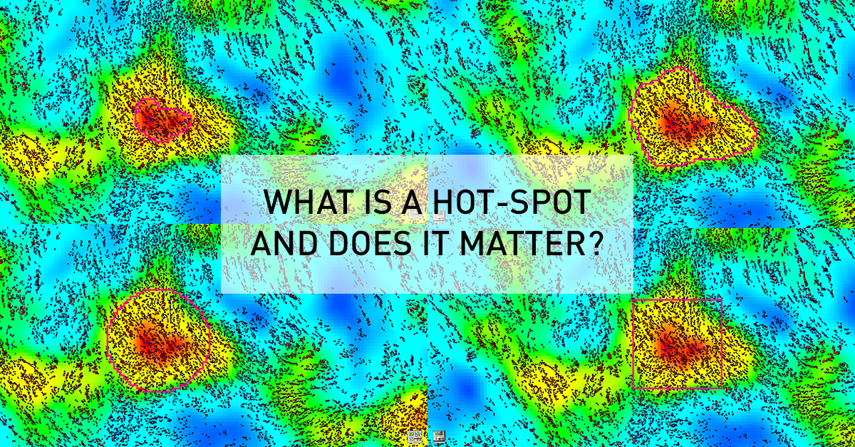
    As an example, this general research tool was used to create an APP for visualizing heatmaps for Ki67 expression across entire tissue sections, and determine the location of hot-spots. Although this seems an intuitively appealing approach for managing heterogeneity, it generates new questions relating to the definition of a hot-spot. Clinical guidelines are usually defining a hot-spot as a square of fixed dimensions, or recommend counting a certain number of cells around the hot-spot. For their presentation at the 29th European Congress of Pathology, Omanovic and Schönauwere comparing five different approaches to defining a hot-spot as outlined in the figure below. Note how the hot spot location and area changes between methods, and that heatmap area and count can be defined along the iso-curve of the biomarker response.

    Need for a standardized approach

    In the study above, they found that different definitions as well as area of hot-spots has a statistically significant impact on the proliferation index. Therefore, the fact that different guidelines and hot-spot definitions are adopted in clinical practice and for research purposes, also means that results cannot be compared. Thus, it will difficult to define generally applicable clinical standards (e.g. cut-offs) until there is a consensus on a unified / optimal definition of a hot-spot.

    Clinical relevance

    Apart from the need for standardization, there is also a need to understand to the impact of the observed differences on the predictive or prognostic power of tissue-based assays. At Karolinska University Hospital, Johan Hartman et. al. are currently exploring this in a study design with a longitudinal cohort, clinical outcomes and access to sequencing data. The outcome of this study will hopefully provide insights allowing for a recommendation of an optimal definition of hot-spots, for understanding the impact on diagnostic/prognostic accuracy, and to what extent a modern tissue-diagnostic approach provide statistically independent information compared to molecular methods. 

    References:

    ​1.Stålhammer et. al.; Digital image analysis outperforms manual biomarker assessment in breast cancer; Modern Pathology 29, 318-329 (2016)

    2.Gudlaugsson et. al.; Comparison of the effect of different techniques for measurement of Ki67 proliferation on reproducibility and prognosis prediction accuracy in breast cancer

    3.KVAST, Swedish breast cancer guidelines, 2018 (in Swedish)

    4.DBCG – Danish Guidelines, May 2017 (in Danish).

  • Managing Tumor Heterogeneity for Quantification of Biomarker Response

    Tumor heterogeneity in research and diagnostics


    Diagnostic pathologists and scientists often rely on tissue data for important diagnostic-, research-, or even business- decisions. One of the major concerns relate to tumor heterogeneity in the context of quantifying tissue biomarker response. Questions revolve around how to reliably visualize, quantify, and in practical terms deal with heterogeneity.

    The impact of heterogeneity on diagnostic accuracy​


    Concerns are apparently well founded. In a recent publication, Stålhammer et. al. [1] demonstrated that manual stratification of breast cancer patients into Luminal A vs. Luminal B based on Ki67 was associated with an error rate of 31%. Using image analysis with computer identified hot-spots reduced the error rate to 19%, when using PAM50 as a pseudo gold-standard.

    Heterogeneity and hot-spots


    What really made the difference in reducing error rates, was automated identification of hot-spots, using image analysis. Gudlagsson et. al [2] showed that as much as 50% of pathologists were unable to correctly identify the hottest hot-spot which, in some cases, can represent a major cognitive challenge. This challenge was effectively mitigated using image analysis.

    Diagnostic applications


    As a first practical diagnostic application, we considered automated hot-spot identification for Ki67 expression in breast cancer. By creating heat maps of biomarker expression, the intended use of the APP is to support pathologists in both visualizing heterogeneity and locate the hottest hot-spot (s). This can be used as input to APPs that quantify biomarker expression in the hot-spot(s). CE-marking for In-Vitro Diagnostic purposes, required special attention to the study designs for validating clinical performance.

    Research applications


    The challenges related to heterogeneity may well be further amplified when interrogating far more complex, and sometimes multiplexed, biomarkers across the entire tumor micro-environment. With Oncotopix® Author, the ability to visualize and quantify heterogeneity wrt. biomarker response has been generalized for tissue-based cancer research.

    Click here to learn more about the CE-IVD Hot Spot APP.

    Download the Hot Spot brochure

    References​

    1.Stålhammer et. al.; Digital image analysis outperforms manual biomarker assessment in breast cancer; Modern Pathology 29, 318-329 (2016)

    2.Gudlaugsson et. al.; Comparison of the effect of different techniques for measurement of Ki67 proliferation on reproducibility and prognosis prediction accuracy in breast cancer

  • Deep Learning Methods Powered by AI in Tissue Image Analysis

    CIO Applications recently sat down with our founder and CEO Michael Grunkin. The technology print magazine focuses on usage of various tech s

    From tedious manual annotation work to fully automated annotations with a streamlined image analysis workflow. This is now possible with AI tools that are remarkably accurate generating powerful results within tissue segmentation, cell identification, quantification and other cell-based tasks.


    AI expands the possibilities in cancer research and diagnostics


    Whether you are looking for nuclei quantification in IHC images, accurate tumor separation in H&E, or advanced context aware region mapping in brain images, Oncotopix®/Biotopix™ AI image analysis is your go-to toolbox. Deep neural networks bring virtually endless possibilities to augmented pathology.

    AI made easy

    The Oncotopix®/Biotopix™ AI platform is train-by-example image analysis for digital pathology, made easy-to-use. Deep learning convolutional neural algorithms are trained based on easily annotated training data provided by you as the user. AI is not magic – you are in control of the learned behavior.

    Patented virtual double stain (VDS)

    Are you already using Artificial Intelligence (AI) for image analysis in digital pathology, and have you experienced the time-consuming task of annotating enough training data to capture the variance in your images? Let Visiopharm introduce you to our unique patented Virtual Double Stain (VDS) method for automatic annotation of training data.

    AI redefines what is possible in image analysis

    In more advanced cases robust identification of morphologically and/or functionally distinct features in histological samples, such as glomeruli in the kidney, compartments of the brain, and tumor/stroma separation, continues to challenge traditional image analysis methods. Differences in disease severity; preanalytical variables, such as variance in staining intensity; and structures or regions reliant solely on context often result in tedious manual annotations to achieve necessary accuracy.

    Stefan Hamann, PhD from the Translational Pathology Laboratory at Biogen has utilized Visiopharm’s context-based deep learning algorithms to automate the laborious manual tissue annotation process. The task involved sagittal mouse brain sections with 6 regions-of-interest. The result is a fully automated mouse brain annotation tool that is remarkably accurate when confronted with unique tissue artifacts, illuminating the possibilities of deep learning within a streamlined image analysis workflow.

    Visiopharm’s AI webinar

    Watch our webinar to experience Oncotopix®/Biotopix™ AI image analysis with real-life applications, where Visiopharm’s deep learning experts demonstrate the “how to” with explanations and relevant demo cases.

    Visiopharm AI image analysis solution


    The Oncotopix® and Biotopix™ AI image analysis modules are available and fully integrated with the Visiopharm current image analysis software platform. Visit visiopharm.com/ai-deeplearning

    Visiopharm AI Image Analysis in the news

    Please see the launch news at TissuePathology: Keith Kaplans Digital Pathology Blog